

A groundbreaking 2026 analysis published in Glia has identified a conserved microglial transcriptional program that is shared across five distinct neurodegenerative contexts: Amyotrophic Lateral Sclerosis (ALS), frontotemporal dementia (FTD), Alzheimer’s disease (AD), aging, and Parkinson’s disease (PD). This significant discovery suggests that despite the diverse clinical presentations and underlying pathologies of these devastating conditions, the brain’s resident immune cells, microglia, may adopt a common reactive state. The most prominent marker associated with this shared program is SPP1, a gene encoding the protein osteopontin, signaling a crucial step towards understanding the universal inflammatory responses in neurodegeneration. However, researchers emphasize that this finding illuminates a cellular-state signal rather than offering a ready diagnostic test, underscoring its primary value as a research-facing insight.
The Evolving Understanding of Microglia in Brain Health and Disease
For decades, microglia were largely considered passive support cells within the central nervous system, primarily tasked with immune surveillance and debris clearance. This perspective has dramatically shifted over the past two decades, with accumulating evidence positioning microglia as dynamic, multifaceted cells critical for brain development, plasticity, and maintaining homeostasis. They constantly survey their environment, prune synapses, respond rapidly to injury or infection, and play a pivotal role in the pathogenesis of various neurological disorders.
In the context of neurodegenerative diseases, microglia’s role becomes particularly complex and often paradoxical. While their acute activation can be beneficial, clearing misfolded proteins and cellular debris, chronic or dysregulated microglial activity is increasingly implicated in exacerbating neuronal damage and disease progression. This dual nature – protective in some contexts, detrimental in others – has made understanding specific microglial states a central challenge in neuroscience. The ability of microglia to shift into various reactive states, some beneficial and some harmful, necessitates precise tools to characterize these states at a molecular level.
Advanced Genomics Unveils Cellular Specificity
The study by Palma et al. leveraged single-nucleus RNA sequencing (snRNA-seq), a powerful technique that measures gene expression in individual cell nuclei. This approach is instrumental for deciphering the heterogeneous cellular landscape of complex tissues like the brain. Unlike bulk tissue RNA sequencing, which averages gene expression across all cell types, snRNA-seq allows researchers to isolate and analyze the unique transcriptional profiles of specific cell populations, such as microglia. This resolution is critical because distinct cell types, even within the same tissue, can respond to disease in vastly different ways, and averaging these signals can obscure important biological insights.
The researchers’ core question was not whether all neurodegenerative diseases are fundamentally the same, but rather if they share a common microglial state. This distinction is vital for clinical translation. Recognizing shared inflammatory programs allows for the development of targeted therapies that might modulate these common pathways, even while acknowledging that disease-specific triggers, protein pathologies (e.g., amyloid-beta in AD, alpha-synuclein in PD), and anatomical vulnerabilities (e.g., motor neurons in ALS, frontal/temporal lobes in FTD) remain distinct.
Methodology: An Integrated Analysis Across Diverse Datasets
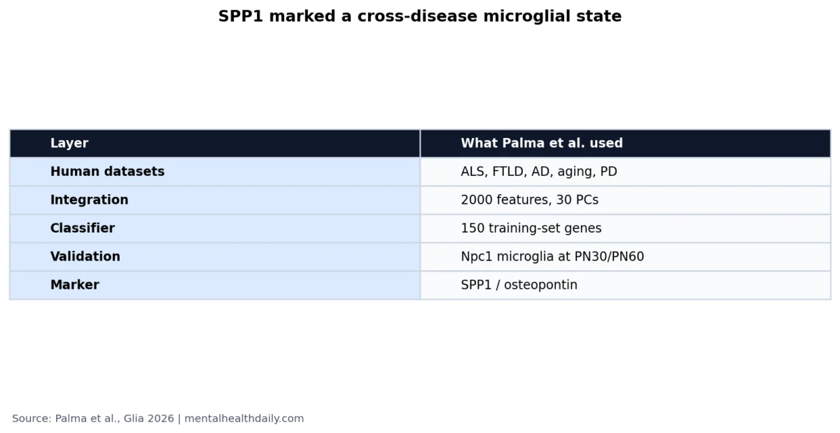
To identify conserved microglial programs, the Palma et al. study meticulously integrated human snRNA-seq datasets sourced from post-mortem brain tissues of individuals affected by ALS, frontotemporal dementia, Alzheimer’s disease, Parkinson’s disease, and those undergoing normal aging. This multi-disease approach is a strength, allowing for the detection of truly universal responses rather than disease-specific quirks.
The analytical workflow was robust, designed to minimize noise and maximize biological signal. After initial data preprocessing, the researchers focused on 2,000 highly variable genes and utilized 30 principal components. Principal component analysis (PCA) is a statistical method that compresses high-dimensional data, like thousands of gene expression levels, into a smaller set of composite variables (principal components) that capture the most significant variation. Using 30 components allowed for a comprehensive representation of gene-expression signals without treating every single gene as equally informative or prone to noise.
A critical step in integrating diverse datasets, which can vary significantly due to different tissue sources, disease stages, laboratory protocols, and sequencing chemistries, was the application of Harmony integration. This algorithm helps to correct for batch effects and technical variations, allowing for a more accurate comparison of cellular states across disparate studies. Following integration, marker-based cluster annotation was used to identify and refine microglial populations, ensuring that the subsequent analysis focused specifically on these immune cells. Visualization techniques such as Uniform Manifold Approximation and Projection (UMAP) were employed to display the structural relationships between cells with similar expression profiles, helping to illustrate the shared patterns identified.
The Five Neurodegenerative Contexts: A Spectrum of Disease
The inclusion of five distinct neurodegenerative contexts underscores the breadth of the findings.
- Amyotrophic Lateral Sclerosis (ALS) is a rapidly progressive and fatal neurodegenerative disease that primarily affects motor neurons in the brain and spinal cord, leading to muscle weakness, paralysis, and ultimately respiratory failure.
- Frontotemporal Dementia (FTD) encompasses a group of disorders characterized by progressive damage to the frontal and temporal lobes of the brain, resulting in profound changes in personality, behavior, language, and executive function.
- Alzheimer’s Disease (AD) is the most common cause of dementia, defined by the accumulation of amyloid plaques and tau tangles, leading to memory loss and cognitive decline.
- Parkinson’s Disease (PD) is a progressive disorder of the nervous system that primarily affects movement, characterized by the degeneration of dopaminergic neurons in the substantia nigra and the presence of Lewy bodies (alpha-synuclein aggregates).
- Aging, while not a disease itself, is the primary risk factor for most neurodegenerative conditions and is associated with significant changes in brain physiology, including chronic low-grade inflammation.
The diversity of these conditions, with their unique clinical symptoms, hallmark protein pathologies, and affected brain regions, makes the discovery of a shared microglial program particularly compelling. It points towards a convergent immune response that transcends the initial disease triggers.
SPP1: A Prominent Marker of the Shared Microglial Program
At the heart of the identified shared microglial program is the gene SPP1, which encodes osteopontin. Osteopontin is a highly versatile secreted glycoprotein known for its roles in cell adhesion, migration, immune cell signaling, and tissue remodeling. In the brain, SPP1 is frequently regarded as a marker of reactive or disease-associated microglial states rather than a direct causative agent of disease. Its upregulation signifies a microglial response that is part of a broader inflammatory and neurodegenerative cascade.
The study’s emphasis on SPP1 was further strengthened by a crucial validation step: the expression of Spp1 was confirmed in primary microglia isolated from a Niemann-Pick type C (NPC) mouse model. Niemann-Pick type C is a rare, severe lipid-storage neurodegenerative disorder that, in mouse models, faithfully recapitulates key features of microglial activation, lysosomal dysfunction, and progressive neurological decline. This biological anchor, demonstrating that the computational signal derived from human data can be observed and localized in an in vivo disease model, significantly bolsters the plausibility of SPP1 as a relevant biological marker. This validation is critical in machine learning-driven analyses, where statistical patterns sometimes lack clear biological counterparts.
The dual nature of osteopontin’s biology – its involvement in both beneficial processes like phagocytic clearance of debris and potentially harmful chronic inflammatory activation – highlights why an "SPP1-positive" state should not be oversimplified as inherently "good" or "bad." Microglial states are dynamic and context-dependent; a particular state might be protective at one stage of a disease or in one brain region, yet detrimental in another. The paper identifies a conserved state but refrains from assigning a universal clinical meaning, encouraging nuanced interpretation.
Implications for Research and the Path to Therapeutic Discovery
Beyond identifying a shared microglial state and its marker, the study explored the potential for computational classification. A random-forest model, trained on the 150 most variable genes from the integrated dataset, demonstrated the ability to classify disease conditions from controls in held-out cells. Random forest models are ensemble machine-learning techniques that combine multiple decision trees, providing robust classification capabilities without necessarily identifying direct causal mechanisms.
This classification capability is a powerful research tool. It indicates that microglial disease states contain sufficient information to be computationally recognized, even if each disease does not possess a perfectly unique microglial pattern. The combination of shared and disease-weighted components can collectively carry a strong signal. Genes repeatedly identified as important for classification, such as SPP1, become strong candidates for further mechanistic investigation in animal models, organoids, or human post-mortem validation studies. For instance, prior work in Alzheimer’s models has already linked SPP1 to microglial phagocytosis and synaptic engulfment, providing existing context for its role.
Caution Against Immediate Diagnostic Application
It is imperative to underscore the "important limit" articulated by the researchers: classifying disease labels from purified microglial transcriptomes in post-mortem brain tissue is fundamentally different from diagnosing a living patient. The complex process involving brain tissue acquisition, single-cell sequencing, dataset harmonization, and curated cell selection is far from a routine clinical assay. Therefore, the finding of an SPP1-positive microglial state is primarily a research-facing implication.
The more significant impact lies in its potential to guide future research and therapeutic development. The identification of a conserved SPP1-positive microglial state provides a valuable cross-disease readout for evaluating whether experimental interventions can modulate or redirect neurodegeneration-associated inflammation. This moves beyond vague notions of "inflammation being bad" to a more specific, molecularly defined target.
Connecting with Prior Research on Disease-Associated Microglia
This new analysis builds upon and extends previous seminal work in the field of neuroinflammation. Studies by Deczkowska et al. (2018), for instance, introduced the concept of "disease-associated microglia" (DAM) as a recurring immune response observed across various neurodegenerative conditions. The Palma et al. study significantly advances this idea by providing integrated human transcriptomics and explicit cross-condition classification, offering a more comprehensive and directly human-relevant validation of the DAM concept.
Furthermore, research by De Schepper et al. (2017) had already linked SPP1 to specific microglial phagocytic states and synaptic engulfment in Alzheimer’s model systems. This prior work contextualizes the SPP1 finding, positioning it not as an arbitrary marker but as part of a well-established axis of reactive microglial responses. Reviews by Bright et al. (2019) on neuroinflammation in frontotemporal dementia have also highlighted how microglia and other immune pathways contribute significantly to disease context, moving beyond purely protein-aggregation narratives. Collectively, this body of literature supports the nuanced thesis that while neurodegenerative diseases are distinct in their etiology and progression, their immune-cell responses can converge on shared pathways.
Evidence Strength, Limitations, and Future Directions
The integrated human single-nucleus datasets provide strong evidence for a conserved microglial transcriptional program across multiple neurodegenerative contexts, with SPP1 emerging as a prominent marker. However, the study explicitly does not support using SPP1 alone as a clinical biomarker, treating SPP1 as universally harmful, or assuming that microglia behave identically across Alzheimer’s, Parkinson’s, ALS, FTLD, and aging. The mouse model validation strengthens biological plausibility but remains preclinical and focused on cell states.
The most effective use of this result lies in informing future neurodegeneration studies. Researchers can now investigate whether a specific therapeutic intervention alters the SPP1-positive state, and critically, whether such a change correlates with improved neuronal survival, synaptic preservation, or other clinically relevant outcomes. The potential to track this state in cerebrospinal fluid or through advanced imaging biomarkers represents a crucial next step, moving the insights closer to clinical applicability. This approach is far more robust than generic claims about "inflammation mattering in brain diseases."
Therapeutic Caution and Biomarker Development
The identification of a shared microglial state does not automatically imply that a single anti-inflammatory drug would be effective across ALS, FTLD, AD, PD, and aging. The timing of intervention, the specific brain region affected, the disease stage, and the surrounding protein pathology all remain critical variables. A therapy that suppresses debris clearance, for example, could be counterproductive if the SPP1-positive state is, in part, a compensatory or beneficial response in certain contexts.
The more promising near-term application is biomarker-guided experimentation. Researchers can design studies to compare whether different disease models converge on the same SPP1-positive program, then rigorously test if modulating this program leads to genuine neuronal improvements or merely shifts a marker without clinical benefit. This distinction is vital for separating a useful therapeutic target from a mere disease-state label.
Furthermore, human validation will require careful consideration of spatial context. A microglial transcriptomic state observed near amyloid plaques, degenerating motor pathways, or vulnerable frontal networks may carry different implications than the same marker found in less affected tissue. Combining single-cell data with spatial transcriptomics and detailed neuropathological analyses will be crucial to fully interpret the SPP1 signal and understand its localized significance. This spatial mapping would also help differentiate a localized injury response from a more widespread, brain-wide inflammatory state.
Such refined analyses would also aid in interpreting negative findings. If, for example, future studies fail to detect SPP1 enrichment in blood or cerebrospinal fluid, it may not signify biological irrelevance but rather that the marker is too tissue-localized for peripheral testing, highlighting the importance of assay choice. Single-nucleus RNA sequencing captures cell-state differences that bulk tissue analysis, serum cytokines, or routine inflammatory markers might miss. Therefore, a weak peripheral SPP1 result would not necessarily contradict a strong microglial signal within degenerating brain tissue; it would merely indicate a difference in the measurement layer.
Ultimately, the safest interpretation of this groundbreaking article is translational, not diagnostic. SPP1 helps to name and characterize a conserved disease-state program within microglia, providing a powerful research tool. Whether future, less invasive assays can capture this signal outside of brain tissue, thereby translating it into a diagnostic or prognostic biomarker, remains an open and exciting question for continued scientific inquiry.
Immediate Questions and Their Answers:
- Is SPP1 a blood biomarker for dementia or Parkinson’s disease? Not based on this paper. The signal was identified through microglial transcriptomics in brain tissue and validated in mouse microglia. A blood biomarker would necessitate independent assay development and rigorous clinical validation in human patients.
- Does shared microglial activation mean these diseases have a single cause? No. Shared immune-cell states can manifest downstream of diverse initial disease triggers, which include the accumulation of amyloid-beta, tau, or alpha-synuclein proteins, motor neuron injury, lysosomal stress, or age-related tissue damage.
- How can researchers utilize the SPP1 signal now? The most immediate and impactful use is for research stratification. SPP1-positive microglial states can serve as a valuable reference point to compare various disease models and to test whether experimental treatments effectively alter a conserved neuroinflammatory program, thereby providing a more precise target for drug development.

