

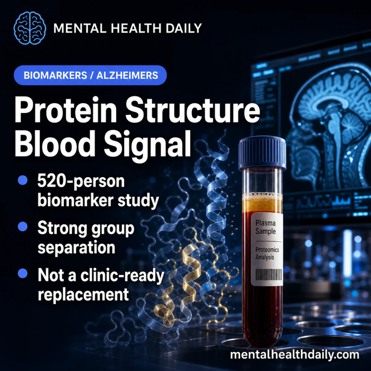
A landmark 2026 study published in Nature Aging, involving 520 participants, has revealed that specific plasma protein-structure signatures can classify healthy controls, individuals with mild cognitive impairment (MCI), and those with Alzheimer’s disease (AD) with a notable 83.44% three-way accuracy. This discovery represents a significant biomarker signal, underscoring the potential for a new dimension in blood-based Alzheimer’s testing, though researchers and clinicians alike emphasize it is not yet a clinic-ready replacement for established diagnostic methods. The findings open new avenues for understanding the complex pathology of Alzheimer’s beyond traditional amyloid and tau markers, potentially offering insights into inflammatory, lipid, and vascular contributions to neurodegeneration.
The Urgent Need for Early and Accurate Alzheimer’s Diagnosis
Alzheimer’s disease, a progressive neurodegenerative disorder, affects millions worldwide, representing the most common cause of dementia. With an aging global population, the prevalence and economic burden of AD are projected to soar, making the development of early and accurate diagnostic tools a critical public health imperative. Current diagnostic methods often involve expensive and invasive procedures such as cerebrospinal fluid (CSF) analysis and positron emission tomography (PET) scans, which measure amyloid plaques and tau tangles in the brain. While highly accurate, these methods are not universally accessible, can be costly, and often employed only when cognitive symptoms are already apparent. Clinical assessment, including neuropsychological testing, remains foundational but can be subjective and may not differentiate AD from other forms of dementia or age-related cognitive decline effectively in early stages.
The challenge lies in detecting Alzheimer’s pathology at its earliest stages, particularly during the mild cognitive impairment (MCI) phase, where interventions might be most effective. MCI is characterized by a measurable decline in cognitive abilities—such as memory, language, or judgment—that is greater than expected for a person’s age but not severe enough to interfere significantly with daily life, thus not meeting the full criteria for dementia. This boundary is clinically crucial because biomarker-guided interventions, including potential disease-modifying therapies, are believed to yield the greatest benefit before irreversible neuronal damage and functional decline become obvious. The ability to accurately identify individuals at the MCI stage who are likely to progress to AD would revolutionize clinical trials, patient stratification, and potentially, future therapeutic strategies.
Unveiling Disease Signatures Through Plasma Structural Proteomics
The innovative approach employed by Son et al. focuses on "plasma structural proteomics," a sophisticated technique that moves beyond merely measuring the concentration of proteins in blood. Instead, it investigates subtle yet crucial changes in protein accessibility and folding states. Proteins are complex molecules whose biological functions are intimately tied to their three-dimensional structures. Alterations in these structures, even if the overall protein concentration remains stable, can signify underlying pathological processes. In the context of Alzheimer’s disease, such structural clues in blood plasma could reflect systemic changes associated with neurodegeneration, inflammation, or metabolic dysregulation.
The methodology involves a chemical-labeling assay known as covalent protein profiling, which tags accessible peptide regions within plasma proteins. These tags provide information about how proteins are folded and which parts are exposed or hidden. By analyzing these structural signatures across a large cohort, researchers can identify patterns indicative of different disease states. This technique offers a unique window into protein function and interaction, distinct from the more common concentration-based blood tests that are currently being developed for Alzheimer’s, such as those measuring specific levels of amyloid-beta (Aβ) peptides or phosphorylated tau (p-tau).
A Deep Dive into the Study’s Findings: Accuracy and Discrimination
The Son et al. study, a significant advancement in the field, utilized this covalent protein profiling to label accessible peptide regions in plasma proteins from 520 participants. Subsequently, machine-learning classifiers were trained to distinguish between three distinct groups: healthy controls, individuals with mild cognitive impairment (MCI), and patients diagnosed with Alzheimer’s disease. The final reported classifier achieved an impressive 83.44% accuracy across these three groups.
This "three-way accuracy" is particularly noteworthy. Classifying between three distinct stages—healthy, MCI, and AD—is inherently more challenging than a simpler binary comparison (e.g., AD vs. healthy controls) because MCI represents a transitional state with overlapping features. The ability of the model to accurately parse these stages suggests a robust underlying biological signal. Beyond overall accuracy, the study also reported high pairwise discrimination rates, quantified by the Area Under the Receiver Operating Characteristic (AUROC) curve. An AUROC of 0.9343 was observed for distinguishing healthy controls from MCI, and an AUROC of 0.9325 for discriminating MCI from Alzheimer’s disease.
AUROC is a widely accepted metric in biomarker development, summarizing how well a test separates different groups across various diagnostic thresholds. An AUROC value near 0.5 indicates a chance-level performance, meaning the test is no better than random guessing. Conversely, values above 0.9 are generally considered strong indicators of a test’s discriminatory power in development datasets. The high AUROC values reported in this study, exceeding 0.93 for both key comparisons, signal a powerful ability to differentiate these clinically relevant groups, positioning this approach as a serious contender in the quest for advanced Alzheimer’s biomarkers.
However, the phrase "development dataset" is crucial. Biomarker models often perform optimally within the specific dataset from which they were derived. The real-world complexity of memory clinics presents a much "messier population," encompassing conditions like vascular cognitive impairment, Lewy body disease, depression, sleep apnea, medication effects, traumatic brain injury, and mixed pathologies, in addition to normal aging. A clinically useful blood test must demonstrate its robustness and maintain high accuracy in such diverse and heterogeneous populations. Nevertheless, these results are far from noise; they set a high bar for the next phase of validation, indicating a signal large enough to warrant extensive replication and comparison with established blood biomarker work. The ultimate utility will also hinge on whether this panel provides incremental value beyond existing clinical data, including age, APOE genotype, plasma p-tau217, amyloid-beta ratios, cognitive screening, and comprehensive clinical history.
Biological Plausibility: Connecting Structural Changes to Alzheimer’s Pathology
The identified panel of structural changes centered on specific peptides from three key proteins: C1QA, Clusterin (also known as apolipoprotein J), and ApoB. The biological plausibility of these proteins’ involvement in neurodegeneration lends significant credibility to the findings, ensuring the classifier isn’t merely a "black-box lottery ticket" but grounded in known disease mechanisms.
-
C1QA: A Link to Complement Biology and Neuroinflammation
C1QA is a component of the C1 complex, the initiating molecule of the classical complement pathway, a crucial part of the innate immune system. The complement system is involved in tagging pathogens and cellular debris for clearance, but its dysregulation has been strongly implicated in neurodegenerative diseases, including Alzheimer’s. In AD, complement activation can contribute to synaptic pruning (an excessive removal of synapses), glial cell activation, and inflammatory signaling, all of which are central to brain pathology. A structural signal involving C1QA in plasma suggests a systemic perturbation in complement biology that could indirectly reflect ongoing brain pathology. -
Clusterin (Apolipoprotein J): A Multifaceted Player in Amyloid and Lipid Homeostasis
Clusterin has a long-standing association with Alzheimer’s disease, appearing repeatedly in genetic studies and biomarker discussions. It is a glycoprotein involved in various biological processes, including lipid transport, inhibition of amyloid aggregation, cellular apoptosis, and response to inflammatory stress. Structural changes in clusterin could indicate altered chaperone activity, impaired lipid handling, or modified interactions with amyloid-beta, offering a distinct pathological insight compared to a simple change in its concentration. Its role in amyloid clearance and its genetic links to AD make it a highly plausible candidate for a disease-modifying biomarker. -
ApoB: Connecting to Lipid Transport and Vascular Risk
ApoB (Apolipoprotein B) is a major structural protein of various lipoproteins, including low-density lipoproteins (LDL), very-low-density lipoproteins (VLDL), and intermediate-density lipoproteins (IDL). These lipoproteins are critical for lipid transport throughout the body. The connection of ApoB to the panel links the structural proteomics findings to lipid metabolism and vascular risk biology. It is increasingly recognized that dementia risk is not solely neurodegenerative; vascular factors play a significant, often co-occurring, role. Patients with Alzheimer’s frequently exhibit mixed pathologies, including amyloid plaques, tau tangles, vascular injury, inflammation, and metabolic risk factors. A structural change in ApoB could reflect systemic lipid dysregulation or vascular compromise that contributes to cognitive decline.
The study also reported a broad accessibility gradient across 879 labeled peptides, with average accessibility decreasing from healthy controls (93.2%) to MCI (92.0%) and further to Alzheimer’s disease (91.1%). This consistent directional shift suggests a global structural-protein alteration across the measured peptide set, hinting at widespread changes in protein folding and accessibility as the disease progresses.
Protein Structure: A New Layer Beyond p-Tau217 and Amyloid Markers
The landscape of Alzheimer’s blood testing has been predominantly shaped by amyloid and tau markers, which align with the ATN (Amyloid, Tau, Neurodegeneration) framework. This framework helps to classify a clinical syndrome by its underlying biological pathology, distinguishing amyloid pathology (A), tau pathology (T), and evidence of neurodegeneration (N). Plasma p-tau217, in particular, has emerged as one of the strongest blood-based candidates, demonstrating remarkable accuracy in tracking Alzheimer’s tau pathology and effectively differentiating AD from other non-Alzheimer’s conditions.
The Son study, however, poses a different, complementary question: Can plasma protein structure provide a parallel signal indicative of disease stage? The answer, within this specific dataset, was affirmative. This new approach does not aim to replace existing amyloid or tau markers but rather to add another layer of information. The most crucial next step involves a direct comparison: does this new protein-structure classifier provide additional, independent information beyond what is already captured by p-tau217, amyloid-beta ratios (e.g., Aβ42/40), neurofilament light (NfL), APOE genotype, age, sex, and standard cognitive testing?
Plasma p-tau217 serves as the obvious benchmark, given its proximity to clinical implementation in various settings. A protein-structure panel could carve out a distinct role if it demonstrates superior accuracy in early MCI classification, identifies specific inflammatory or lipid-dominant subgroups of AD, or helps clarify diagnoses for individuals whose standard amyloid/tau markers are borderline or inconclusive.
Beyond diagnosis, another potential application is disease staging. If structural accessibility consistently shifts from healthy control to MCI to Alzheimer’s disease, longitudinal studies could investigate whether these panel changes correlate with symptom progression over time. The study’s small longitudinal subset, though preliminary, showed encouraging directional trends with 86.0% classification accuracy, hinting at its potential for monitoring disease trajectory.
Interpreting APOE Genotype Signals and the Challenge of Personalized Medicine
The study also revealed that the C1QA signal varied by APOE genotype, with APOE epsilon4/epsilon4 carriers exhibiting the lowest accessibility. The APOE epsilon4 allele is the strongest common genetic risk factor for late-onset Alzheimer’s disease. However, it is crucial to interpret this finding with caution. Carrying one or two copies of epsilon4 increases risk but is not a deterministic verdict; many carriers never develop dementia, and a significant proportion of Alzheimer’s cases do not carry the epsilon4 allele.
This genotype-dependent signal is intriguing because it suggests that the structural-protein panel might partly capture pathways linked to genetic risk, complement activation, or lipid biology, which are known to be modulated by APOE. However, such a finding cannot be treated as a definitive genetic-risk verdict without extensive prospective outcome data demonstrating its predictive power in diverse populations. It underscores the complexity of AD and the need for personalized diagnostic approaches that integrate genetic, biomarker, and clinical information.
The Real Test: External Validation and Clinical Utility
This study, while groundbreaking, is fundamentally a biomarker-development study. It effectively demonstrates that protein-structure signatures can differentiate groups within the analyzed cohorts. However, it cannot yet prove that this panel will maintain the same level of accuracy and utility in primary care settings, specialized memory clinics, or in populations with mixed dementia, depression-related cognitive complaints, vascular cognitive impairment, Parkinson’s disease dementia, or across diverse racial and socioeconomic backgrounds.
The challenge of "external validation" is paramount. Blood biomarkers, despite their promise, carry a "false-precision hazard." If a highly accurate test is applied to the wrong population—for instance, a general population with a low prevalence of AD—even high sensitivity and specificity can generate a substantial number of false positives, leading to unnecessary anxiety, further costly investigations, and potential misdiagnosis. Therefore, the most pragmatic initial clinical use of such a test would likely be as a triage tool: identifying individuals who warrant further confirmatory amyloid/tau testing, specialist neurological review, or longitudinal follow-up.
External validation studies must report more than just overall accuracy. They need to meticulously detail performance metrics across various demographic and clinical factors, including age, sex, APOE genotype, race, kidney function, inflammatory disease status, vascular disease, depression, and medication exposure. These factors can significantly influence plasma protein profiles and could potentially make a structural-proteomics assay appear more robust in carefully controlled research cohorts than it would in routine clinical practice.
Furthermore, the test must be calibrated against outcomes that are truly meaningful to patients and clinicians: conversion rates from MCI to dementia, rates of cognitive decline, future amyloid/tau positivity as confirmed by gold-standard methods, and the ability to definitively distinguish Alzheimer’s disease from other non-Alzheimer’s dementias. While high AUROC values are undoubtedly promising, demonstrating superior longitudinal clinical discrimination remains the more challenging and ultimately more impactful target.
The practical workflow of such a test is another critical consideration. An analytically excellent blood test can be rendered impractical if the subsequent clinical steps are unclear. A positive plasma protein-structure result should ideally trigger confirmatory amyloid/tau testing or specialist review, rather than an immediate, definitive dementia label. Conversely, a negative result should be interpreted within the broader clinical context, considering symptoms, age, family history, medication burden, sleep patterns, depression, and other vascular risk factors.
The Son et al. study significantly expands the blood-biomarker conversation beyond the established amyloid and tau markers. If these protein-structure signatures are independently replicated and validated, they could become a complementary diagnostic layer, offering deeper insights into inflammatory, lipid, or complement biology in patients whose standard Alzheimer’s markers might only tell part of their complex story.
Future Directions: Strict Comparator Design and Personalized Approaches
For this promising research to translate into clinical utility, the next generation of studies must employ strict comparator designs. This means testing the structural proteomics panel head-to-head against plasma p-tau217, amyloid-beta ratios, neurofilament light, APOE genotype, and a comprehensive set of ordinary clinical variables, all within the same patient cohorts. A new assay earns its place in clinical practice only if it demonstrably improves classification accuracy, disease staging, or prognostic prediction beyond what these existing and often easier-to-interpret signals already provide.
The journey from a compelling research finding to a clinically validated and implemented diagnostic tool is arduous and typically spans years. However, the discovery of robust plasma protein-structure signatures represents a vital step forward. It underscores the potential of advanced proteomics to uncover novel biological pathways involved in Alzheimer’s disease and offers hope for a future where early, accurate, and accessible blood tests can revolutionize the diagnosis and management of this devastating condition, paving the way for more targeted and effective interventions.

