

A landmark 2026 adolescent depression study, utilizing resting-state functional magnetic resonance imaging (fMRI), has offered compelling evidence that childhood trauma imprints a measurable network signature within the brains of depressed teenagers. Researchers identified significant disruptions in the default-mode network (DMN) hubs among adolescents diagnosed with major depressive disorder (MDD) who also reported histories of childhood trauma. Crucially, the study also demonstrated that baseline functional-connectome patterns could predict treatment response with an impressive 82% accuracy, marking a significant stride towards personalized mental health care for a vulnerable population.
This extensive research, which included 492 adolescents, stands out for its robust sample size in the often-underpowered field of psychiatric imaging, particularly concerning the adolescent demographic. The findings not only deepen the understanding of the neurobiological underpinnings of trauma-informed depression but also pave the way for potential future diagnostic and prognostic tools, albeit with necessary caveats regarding clinical readiness.
The Silent Scars: Understanding Childhood Trauma and Adolescent Depression
Adolescent depression is a pervasive global health challenge, affecting millions of young people during a critical developmental period. The World Health Organization estimates that depression is a leading cause of illness and disability among adolescents worldwide. When compounded by childhood trauma—experiences such as abuse, neglect, or household dysfunction—the prognosis for recovery often becomes more complex and challenging. Studies consistently show that exposure to early life stress significantly increases the risk for developing MDD, post-traumatic stress disorder (PTSD), and other mental health conditions later in life, often leading to more severe and treatment-resistant forms of these disorders.
The unique vulnerability of adolescents stems from ongoing brain development, particularly in areas governing emotion regulation, executive function, and social cognition. Traumatic experiences during these formative years can profoundly alter neural circuitry, leading to persistent changes in how the brain processes stress, fear, and social information. Despite this established link, the precise neural mechanisms through which childhood trauma influences the trajectory and treatment response of adolescent depression have remained largely elusive, often hindering the development of targeted interventions. This 2026 study directly addresses this gap by seeking a quantifiable neural signature.
Innovating with Neuroimaging: The Role of fMRI and Graph Theory
The study’s methodology leveraged advanced neuroimaging techniques, specifically resting-state fMRI, which measures brain activity by detecting changes in blood flow. Unlike task-based fMRI, resting-state fMRI captures spontaneous fluctuations in brain activity when a person is not performing a specific task, revealing intrinsic functional connectivity—how different brain regions communicate with each other. This approach is particularly valuable for studying complex, network-based disorders like depression, where symptoms often arise from widespread dysfunction rather than isolated lesions.
To interpret the intricate patterns of brain connectivity, the researchers employed graph theory, a powerful mathematical framework used to describe networks. In this context, brain regions are conceptualized as "nodes," and the functional connections between them are "edges." Metrics derived from graph theory, such as efficiency, path length, degree, and small-worldness, provide quantitative measures of how information flows through the brain’s intricate system. For instance, "efficiency" describes how quickly information can travel between nodes, while "small-worldness" indicates a balance between specialized and integrated processing. In depression research, graph metrics are particularly insightful because complex phenomena like mood regulation, memory processing, attention, sleep, and self-referential thinking are known to rely on distributed neural circuits rather than being localized to a single brain area. By analyzing these network properties, scientists can gain a more holistic understanding of how psychiatric conditions alter brain organization.
A Deep Dive into the Study’s Structure and Participants
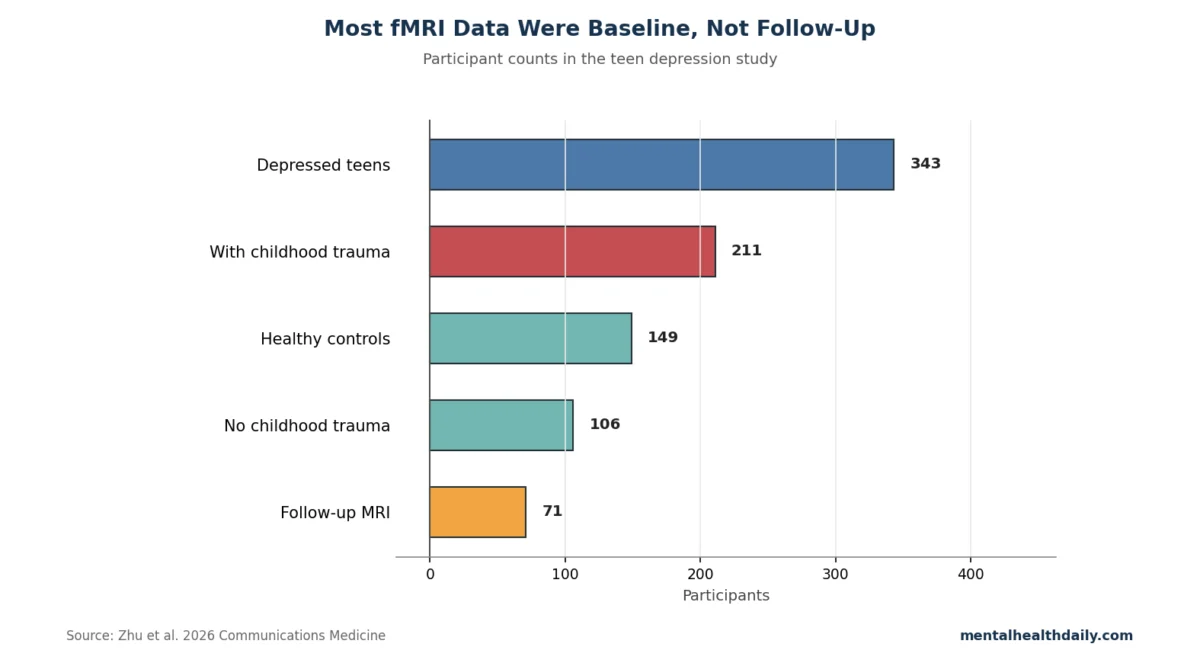
The study, led by Zhu et al., was meticulously designed to capture a comprehensive view of adolescent depression and trauma. It recruited a substantial cohort of 492 adolescents aged 10 to 18 years, a scale that significantly bolsters its statistical power and generalizability compared to many prior psychiatric imaging studies. Within this cohort, 343 participants had been diagnosed with first-episode major depressive disorder, while 149 served as healthy controls.
A critical component of the study was the detailed assessment of childhood trauma. Using established Childhood Trauma Questionnaire cutoffs, researchers identified 211 teenagers within the depressed group who met criteria for exposure to at least one trauma domain. Another 106 depressed teenagers reported no such trauma, and data for 26 participants were unavailable. This careful stratification allowed for direct comparisons between depressed adolescents with and without trauma histories, a crucial step in isolating the specific neural signatures associated with trauma.
While the large overall sample size is a major strength, providing robust data for identifying baseline network differences, the treatment-response component was necessarily smaller. A subgroup of 71 participants returned for follow-up imaging after a period of treatment, with an average treatment duration of 44.22 days. This smaller follow-up group, though limited in size for long-term prediction, still provided valuable insights into the dynamic changes in brain networks in response to therapeutic interventions. The chart depicting participant counts illustrates the scale of the initial recruitment versus the follow-up, emphasizing that the impressive 82% response classifier, derived from baseline connectome data, requires rigorous external validation before it can inform clinical care directly.
Childhood Trauma Imprints on the Default-Mode Network
A central finding of the study was the clear association between childhood trauma and significant disruption within the brain’s default-mode network (DMN). The DMN is a set of interconnected brain regions that are most active when an individual is not focused on the outside world, but rather engaged in internal processes such as self-referential thought, autobiographical memory recall, future planning, and mind-wandering. In adolescent depression, this network is particularly relevant because symptoms like rumination (repetitive negative thinking), self-blame, and threat-biased memory often become intertwined and exaggerated, making the DMN a plausible target for trauma-sensitive alterations.
Zhu et al. reported specific trauma-linked disruptions in key DMN hubs, including the left parahippocampal gyrus, the posterior cingulate gyrus, and the temporal pole. Each of these regions plays a distinct, yet interconnected, role in cognitive and emotional processing:
- Left parahippocampal gyrus: This region is vital for binding memory with its context, helping individuals recall not just an event but also where and when it occurred. Disruption here could contribute to the fragmented or intrusive memories often seen in trauma.
- Posterior cingulate gyrus (PCG): Considered a central hub of the DMN, the PCG is deeply involved in self-referential processing, emotional regulation, and memory retrieval. Its disruption can severely impair an individual’s ability to regulate mood and integrate past experiences.
- Temporal pole: This area contributes significantly to social and emotional meaning, including understanding social cues and processing complex emotions. Alterations here could impact social functioning and the interpretation of emotional stimuli, both of which are commonly affected by trauma.
The researchers emphasize an important interpretive boundary: while the study demonstrates a strong association between childhood trauma, depressive illness, and specific brain-network topology within this sample, it cannot definitively prove that trauma caused these imaging patterns in any individual teenager. The cross-sectional nature of the primary imaging design reveals that these factors "travel together," highlighting a critical area for future longitudinal research to establish causality.
Treatment Response and Network Reorganization
Further enhancing the study’s impact were the findings from the follow-up scan subgroup. These 71 participants, after an average of 44.22 days of treatment, showed encouraging signs of partial normalization in their brain networks, particularly in the left precuneus and amygdala. The precuneus, a medial parietal default-mode hub, is deeply involved in self-related imagery and memory, contributing to self-awareness and consciousness. The amygdala, a well-known subcortical structure, plays a crucial role in assigning emotional salience, especially to threat, reward, and social cues.
Changes in the nodal degree (number of connections) and nodal efficiency (ease of information flow) within the left precuneus were found to correlate significantly with concurrent improvements in both anxiety and depression scores. Interestingly, the study observed biological heterogeneity in response patterns. In one subgroup analysis, participants with less overall symptom change showed a strong negative correlation between the change in nodal efficiency and the change in HAMD-17 (Hamilton Depression Rating Scale) scores (r = −0.634, p < 0.001), a relationship not observed in the high-change subgroup. This suggests that different individuals may achieve clinical improvement through varied neurobiological pathways. Some teenagers might improve through a discernible reorganization of their brain networks, which standard symptom scales might only partially capture. Others might respond primarily to medication, experience improvements through sleep recovery, family stabilization, psychotherapy exposure, or even through regression toward the mean, illustrating the multifaceted nature of recovery.
The Promise and Pitfalls of 82% Prediction Accuracy
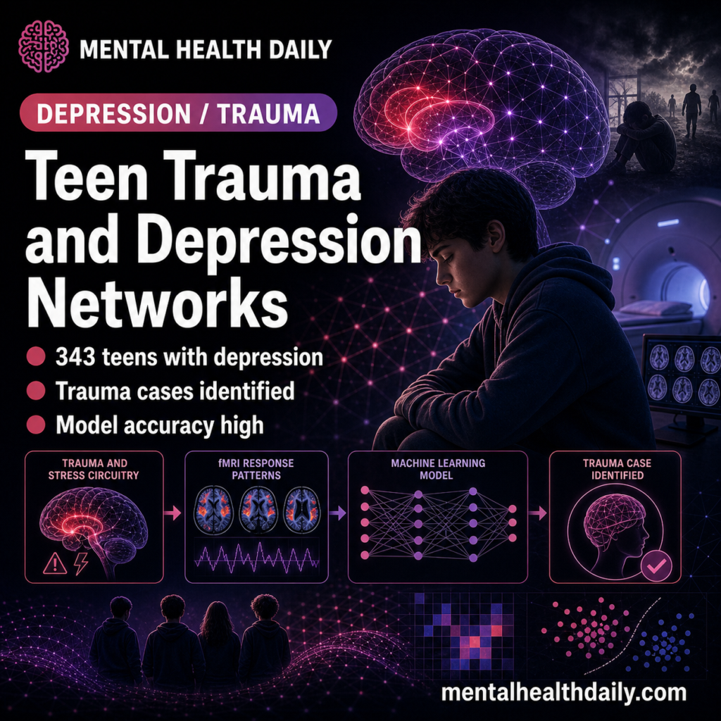
The machine-learning model, which used baseline functional-network matrices to classify treatment responders and non-responders, achieved an impressive 82% accuracy. This level of predictive power is highly significant, especially given the inherent difficulty in predicting treatment outcomes for adolescent depression, a condition known for its variability and heterogeneity. The potential for such a tool to guide clinical decisions, allowing for more precise and individualized treatment plans, is immense.
However, the researchers and independent experts strongly emphasize critical caveats. An 82% accuracy within one imaging cohort is not equivalent to a deployable clinical prediction tool. For a biomarker to be clinically useful, it requires rigorous external validation across a wide range of variables: different fMRI scanners, diverse geographical locations, various treatment types (e.g., specific medications, psychotherapies), varied trauma distributions, different medication protocols, and the presence of comorbid conditions. Without such extensive validation, the model’s accuracy could be specific to the particular scanner, patient population, or treatment context of the original study.
Furthermore, the treatment context itself is crucial. The average follow-up interval of 44.22 days is sufficient to capture early symptom reduction as measured by scales like the HAMD-17, but it is not long enough to confirm durable remission, prevent relapse, facilitate school recovery, or ensure sustained safety. Therefore, the model, in its current form, predicts early response status within this specific research cohort, not full recovery from complex trauma-linked depression. Future studies with longer follow-up periods will be necessary to determine whether these network features can predict relapse, functional recovery, and long-term safety. The research community has seen similar promising initial findings in depression neuroimaging, such as the resting-state connectivity biotypes proposed by Drysdale et al., which later faced concerns regarding stability and reproducibility. This underscores the need for cautious optimism and extensive replication. The adolescent trauma study is best understood as a significant "mechanism-and-prediction lead," offering a vital direction for future research, rather than a finished diagnostic test ready for immediate clinical application.
Translating Research into Clinical Practice: A Roadmap
The 82% prediction number, while exciting, necessitates careful translation for clinical utility. A classifier achieving this accuracy might be identifying genuine treatment-relevant biology, but it could also be capturing scanner-specific quirks, site-specific treatment patterns, or sample characteristics tied to trauma severity and illness burden. For a prediction model to be truly clinically useful, it must answer practical questions before treatment commences. For example, which specific teenager would benefit most from ordinary antidepressant care, trauma-focused psychotherapy, family intervention, sleep treatment, intensive monitoring, or a faster switch to an alternative modality? The Zhu et al. study did not test treatment assignment; rather, the model predicted response within the care pathway already utilized in the study.
Beyond Symptoms: Comprehensive Care for Trauma-Specific Depression
The study’s findings reinforce a critical understanding in mental health: teenagers with depression and childhood trauma often require care that extends far beyond simple mood symptom reduction. The complex interplay of sleep disruption, heightened threat sensitivity, challenging family contexts, school avoidance, dissociative symptoms, elevated self-harm risk, and substance exposure can all perpetuate symptoms, even when a primary medication reduces sadness or anxiety.
Measurement-based care, which involves repeated symptom scales, remains essential. However, for trauma-linked depression, this must be augmented with consistent safety checks, tracking of school function, comprehensive sleep assessment, mapping of family support systems, and vigilant attention to avoidance patterns. While a brain-network marker could eventually add crucial biological context, it would not, and should not, replace the direct measurement of daily life functioning.
The strongest immediate clinical implication from this research is prioritization. Depressed teenagers with documented trauma histories should be considered a higher-risk subgroup and receive faster, more intensive, and more measurement-based follow-up than those with low-risk mild depression. Their potentially distinct network dynamics and lower remission rates necessitate a more proactive and holistic treatment approach.
Guidance for Families: Interpreting the Imaging Signal
For families, the study offers a nuanced, rather than simplistic, message. It is not that a brain scan can definitively "explain" a teenager’s depression in isolation. Instead, the valuable takeaway is that trauma-linked depression can involve measurable, stress-sensitive brain networks, suggesting a biological underpinning that may necessitate more active and comprehensive follow-up than cases of low-risk depression. If a teenager shows partial improvement but continues to struggle with avoidance, sleeplessness, self-harm, or persistent threat reactivity, this imaging signal, combined with clinical observation, reinforces the need for treatment adjustment rather than simply accepting the initial response as sufficient.
The ultimate care target must encompass both symptom remission and improved daily functioning. Practical endpoints such as consistent school attendance, regular sleep patterns, healthy peer contact, reduced family conflict, decreased substance exposure, and a significant reduction in self-harm urges are critical markers of recovery that imaging studies, while informative, cannot replace. These real-world outcomes remain the gold standard for assessing therapeutic success.
Medication Response as One Piece of a Larger Recovery Puzzle
Given the study’s average follow-up period of approximately six weeks, it primarily captured early symptom movement rather than durable, long-term recovery. Teen depression, especially when complicated by trauma, often demands a much longer treatment horizon that includes relapse prevention, restoration of healthy sleep, stabilization of family dynamics, successful school re-entry, and a reduction in avoidance behaviors. A teenager might show a lower HAMD-17 score, indicating symptomatic improvement, yet still remain functionally "stuck" in crucial aspects of their daily life.
The practical endpoint for recovery should always mean fewer symptoms plus better daily function. While imaging biomarkers may eventually help identify risk and guide initial treatment choices, ongoing care must continuously track real-world recovery week by week, ensuring that improvements translate into tangible gains in a teenager’s quality of life and overall well-being.
Addressing Key Questions About Teen Depression, Trauma, and fMRI
The study naturally raises several important questions for the public and clinical communities:
Does this mean childhood trauma permanently changes the brain?
No. The follow-up data from the study provided encouraging evidence of movement toward normalization in some brain regions after treatment. This finding strongly supports the concept of neuroplasticity—the brain’s remarkable ability to reorganize itself by forming new neural connections throughout life—rather than suggesting a fatalistic, permanent alteration. The brain, particularly during adolescence, remains highly adaptable.
Can a teenager get an fMRI to pick an antidepressant?
Based on the current evidence from this study, no. The 82% classifier is a significant research result, indicating a promising area for future development, but it is not yet a clinically validated test. As the researchers emphasized, extensive external validation across diverse clinical settings, patient populations, and treatment types would be absolutely required before such a tool could reliably guide individual patient care decisions, such as antidepressant selection.
Why does trauma lower remission rates in depression?
Childhood trauma introduces a multitude of complex mechanisms that can make standard symptom-only treatment for depression less effective or complete. These include persistent threat learning (where the brain is constantly on alert for danger), chronic sleep disturbance, increased family stress, dissociative symptoms (feeling detached from oneself or reality), pervasive self-blame, and disruptions in attachment patterns. These factors collectively create a more entrenched and multifaceted form of depression that often requires a broader, more integrated therapeutic approach to achieve full and sustained remission.
This 2026 study represents a critical step forward in understanding the intricate relationship between childhood trauma, adolescent depression, and brain network function. Its findings lay crucial groundwork for the development of more personalized and effective interventions, ultimately offering hope for improved outcomes for a generation grappling with the profound impacts of early life adversity.

