

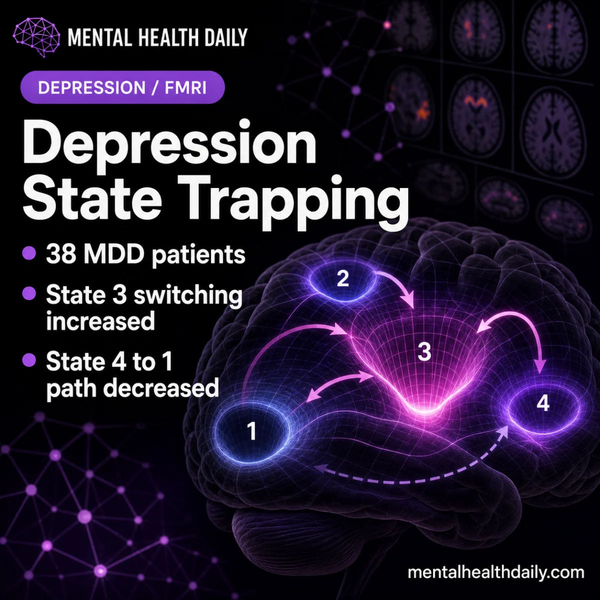
A recent study published in Nature Communications has revealed that individuals with major depressive disorder (MDD) exhibit distinct patterns of brain activity and transitions between these states compared to healthy controls, providing crucial insights into the neurobiological underpinnings of depression. The research, involving a 76-person functional magnetic resonance imaging (fMRI) study, specifically found that MDD patients spent a significantly higher proportion of time in a model-derived "State 3" and showed altered transitions between other states, with these dynamic patterns correlating directly with depression severity and anhedonia. This groundbreaking work points towards altered brain-state transitions as a hallmark of depression, moving beyond static connectivity models to a more dynamic understanding of the disorder.
The study, led by Kilic et al., analyzed resting-state fMRI data from 38 individuals diagnosed with MDD and 38 healthy controls. Resting-state fMRI measures brain activity when a person is not engaged in a specific task, allowing researchers to observe spontaneous fluctuations and identify recurring patterns of neural communication. Rather than focusing on static connections between brain regions, this research employed advanced computational models to cluster brain activity into four distinct, recurring "states." The researchers then meticulously tracked how often participants entered, occupied, and transitioned between these states, providing a temporal dimension to brain function that traditional analyses often miss.
Unpacking State 3: A Window into Depressive Dynamics
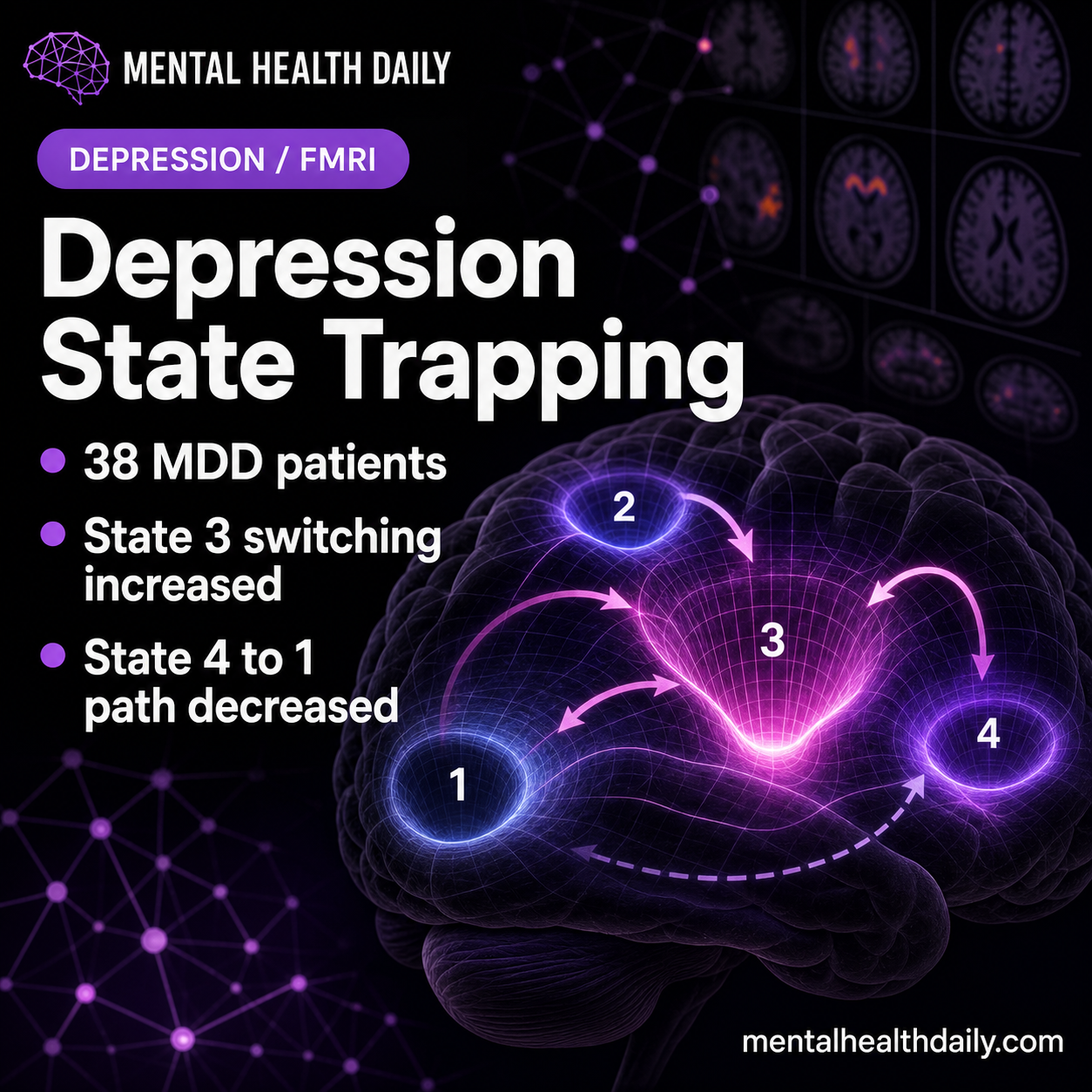
One of the most compelling findings centered on "State 3." MDD participants were found to spend a higher fractional occupancy in this state (27.63% compared to 25.65% in healthy controls, with a statistically significant p-value of 0.035 after False Discovery Rate correction). State 3 was characterized by low-amplitude activity in critical brain regions such as the frontoparietal and default-mode networks, coupled with higher activity in visual and dorsal-attention systems. The frontoparietal network is crucial for cognitive control, attention, and task management, while the default-mode network is active during self-referential thought, introspection, and mind-wandering. The dorsal-attention system, conversely, is involved in externally directed attention.
Intriguingly, while MDD participants occupied State 3 more frequently, they also entered and exited it more often. This pattern suggests that the depressed brain is not simply "trapped" in State 3 in a sustained manner, but rather repeatedly switches into and out of this reactive state. This dynamic instability contrasts sharply with the notion of calm, sustained residence in a particular state. The researchers’ "plain English" interpretation highlighted this nuance: "the depressed brain signal looked more like repeated switching into and out of a reactive state than like calm, sustained residence in one state." This frequent, albeit transient, occupancy of State 3 aligns with clinical observations of cognitive inflexibility, difficulty disengaging from internal rumination, and heightened reactivity to stimuli often seen in depression.
Clinical Relevance: State 3 Occupancy and Symptom Severity
The significance of the State 3 finding was further amplified by its strong correlation with clinical symptoms. Among MDD participants, State 3 fractional occupancy was directly linked to the Quick Inventory of Depressive Symptomatology (QIDS) scores, a widely used scale to quantify depression severity (r² = 0.268, p=0.008 after FDR correction). This established a concrete bridge between an abstract brain dynamic and tangible clinical manifestations.
Moreover, the study delved deeper, connecting the entry into and exit from State 3 to measures of depression and anhedonia. Anhedonia, defined as the reduced ability to experience pleasure or interest, is a core and often debilitating symptom of depression, frequently associated with treatment resistance. The link between State 3 dynamics and anhedonia is particularly meaningful because it connects a specific brain pattern to a central clinical feature, offering a more nuanced understanding than a general correlation with overall depression severity. This dynamic analysis adds a crucial temporal dimension to the established understanding of how default-mode and control networks are implicated in depression, moving beyond static connectivity studies to examine how the brain transitions between different network patterns over time.
Altered Transitions: State 4 to State 1 and General Distress
Beyond State 3, the study identified another critical dynamic alteration: reduced transition probability from "State 4" to "State 1" in MDD patients. Healthy controls showed a mean probability of 0.080 for this transition, while MDD participants exhibited a significantly lower probability of 0.058 (p=0.042 after FDR correction). This particular transition carried substantial clinical meaning due to its directional nature.
Crucially, the clinical model demonstrated a direct inverse relationship: as the State 4 to State 1 transition probability increased, scores for depression, anxiety, rumination, and general distress decreased. The most robust association was observed with general distress, where an r² value of 0.410 (p=0.003 after FDR correction) indicated a strong correlation. This finding is highly interpretable, suggesting that the inability or reduced propensity for the brain to move from State 4 to State 1 is associated with increased psychological burden. This level of specificity is a significant advance over broad statements about "altered connectivity" and offers a clearer target for future research and potential interventions.
Network Control Theory: Bridging Dynamics and Structure
To provide a mechanistic framework for these observed dynamic alterations, the researchers employed "network control theory." This advanced computational approach utilizes the brain’s structural connections (derived from diffusion imaging, which maps white matter tracts) to estimate the energetic cost required for the brain to shift from one activity state to another. In essence, it helps understand how the physical wiring of the brain constrains or facilitates its dynamic functional changes.
The model suggested that certain transitions in MDD followed "more demanding routes," particularly the shift from State 3 to State 2, implying that the depressed brain expends more energy or faces greater structural resistance to make these transitions. Conversely, transitions like State 4 to State 1 appeared "less available," aligning with the observed reduced probability. This integration of functional dynamics with structural constraints provides a deeper, more mechanistic understanding of why brain-state movements might be altered in depression, moving beyond purely descriptive observations of activity patterns. Earlier work in network control theory has consistently demonstrated how structural wiring can profoundly influence and constrain brain-state movement, providing a solid theoretical basis for these interpretations.
Understanding "Brain States": Beyond Anatomical Labels
It is critical to emphasize that "State 1," "State 2," "State 3," and "State 4" are not anatomical regions within the brain. They are recurring activity patterns or configurations of neural networks, computationally generated by clustering fMRI time series data. This distinction is vital for accurate interpretation: the study maps dynamic configurations of known brain networks, rather than proposing four new, localized "depression centers."
The study highlights the interplay of several key brain networks. The default-mode network (DMN) is known for its activity during self-referential thought and internal mentation, often implicated in rumination. The frontoparietal network (FPN) encompasses control regions vital for attention, working memory, and task management. The dorsal-attention network (DAN) supports externally directed attention. Depression research frequently investigates how these systems interact, as symptoms like rumination, anhedonia, and cognitive inflexibility are often linked to abnormal switching or imbalance between internal and external attentional processes. State 3, with its unique mix of low activity in frontoparietal and default-mode regions and higher activity in visual and dorsal-attention systems, represents a complex configuration that, when frequently occupied, correlates with symptomatic burden.
Implications for Research and Future Therapeutic Avenues
This dynamic-state analysis marks a significant evolution in depression research, offering a more nuanced perspective than average connectivity measures. Average connectivity can obscure critical temporal sequences, meaning a depressed and non-depressed brain might exhibit similar average coupling between networks but move through those states in profoundly different ways over time. Transition analysis, by attempting to recover this temporal sequence, provides a richer, more clinically interpretable understanding of brain dysfunction.
The robust connection to anhedonia further enhances the clinical specificity of these findings. Depression is a heterogeneous disorder, encompassing a wide array of symptoms including low mood, rumination, anxiety, sleep disturbances, fatigue, and psychomotor changes. A brain-state result linked specifically to anhedonia—a central feature reflecting loss of reward, interest, and motivation—is far more informative than one tied solely to a total depression score. A brain pattern characterized by repeated entry into reactive or attention-heavy states, coupled with impaired transitions to other states, plausibly maps onto reduced flexibility, persistent rumination, or blunted reward engagement. While the study does not prove a causal chain, it establishes a concrete enough link to guide future investigations.
However, the authors prudently caution that this was a research fMRI and diffusion-imaging analysis, not a treatment trial. It describes depression-linked brain dynamics but cannot yet prove that stimulating a specific region, altering a network, or targeting State 3 would improve symptoms. Clinical translation will require rigorous replication and further research. The findings need to be validated in independent depression cohorts, testing whether the same State 3 and State 4 patterns are consistently observed. Future studies should also account for factors such as medication status, illness duration, patient movement in the scanner, and specific preprocessing choices, all of which can influence resting-state estimates.
Toward Personalized Medicine and Targeted Interventions
Looking ahead, this dynamic approach holds immense promise for personalized medicine. Future work should explore whether patients with different symptom profiles—e.g., melancholic, anxious-ruminative, anhedonic, or inflammation-linked depression—exhibit distinct state-transition patterns. This level of granularity could lead to biomarkers that guide treatment selection.
The ultimate "hard test" for these findings lies in longitudinal treatment studies. If interventions like psychotherapy, medication, sleep optimization, exercise, or neurostimulation successfully reduce anhedonia or other symptoms, researchers must then investigate whether State 3 occupancy and State 4 to State 1 transition probability shift in tandem with symptom improvement. Without this crucial longitudinal step, the current results remain a powerful "state marker" rather than a definitive "treatment target."
In conclusion, the Kilic et al. study represents a significant leap in understanding the dynamic brain in major depressive disorder. By identifying specific alterations in brain state occupancy and transition probabilities, particularly those linked to anhedonia and overall symptom severity, the research provides a sophisticated framework for conceptualizing depression as a disorder of dynamic brain function. While not yet a diagnostic tool or a direct guide for brain stimulation, this work lays essential groundwork for developing future biomarkers and targeted interventions that could revolutionize the diagnosis and treatment of depression, ultimately offering more personalized and effective care for millions worldwide.

