

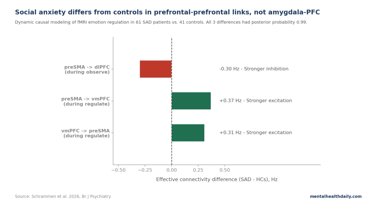
A groundbreaking 2026 study employing advanced brain imaging techniques has significantly reshaped the understanding of social anxiety disorder (SAD), challenging a long-held textbook model of its neural underpinnings. Contrary to the prevailing theory that an overactive amygdala and a deficient prefrontal cortex are at the core of SAD, new research by Schrammen et al. indicates that the critical differences lie not in amygdala-prefrontal connectivity, but predominantly within the intricate network of connections between various prefrontal brain regions, particularly involving the pre-supplementary motor area (preSMA). This discovery, leveraging dynamic causal modeling (DCM), offers a more nuanced view of how the brain processes and regulates social fear, pointing toward novel avenues for research and potentially, future therapeutic interventions.
Challenging a Decades-Old Paradigm
For over two decades, the dominant neural model of social anxiety disorder has been straightforward: an amygdala that hyper-reacts to socially threatening cues, coupled with a prefrontal cortex that fails to adequately inhibit this threat response. This imbalance was widely believed to be the primary driver of clinical symptoms, such as an intense and persistent fear of negative evaluation in social situations. This "broken amygdala-prefrontal regulation" narrative, while intuitive, was largely constructed from functional connectivity studies, which measure statistical correlations in activity between brain regions but cannot definitively ascertain the direction or causality of these influences.
Social anxiety disorder affects approximately 12% of the U.S. population over their lifetime, making it one of the most common anxiety disorders. Its debilitating nature often leads to significant impairment in academic, occupational, and social functioning. Symptoms can range from intense nervousness and avoidance of social gatherings to physical manifestations like blushing, sweating, and trembling. The pursuit of a precise neural signature for SAD has been a significant focus of neuroimaging research, aiming to uncover biomarkers that could inform more effective treatments.
The Power of Directed Connectivity: Dynamic Causal Modeling (DCM)
The pivotal shift in the Schrammen et al. study stems from its use of Dynamic Causal Modeling (DCM), a sophisticated neuroimaging analysis technique that goes beyond mere correlation. While traditional functional connectivity merely identifies whether two brain regions show similar patterns of activity (i.e., they "co-vary"), DCM builds a generative model of neural activity. This allows researchers to estimate directed connectivity, assigning a specific direction (e.g., region A influences region B), a sign (excitatory or inhibitory), and a strength (in hertz) to each connection.
To illustrate, if functional connectivity observes a negative correlation between the amygdala and the ventromedial prefrontal cortex (vmPFC), it cannot tell whether the vmPFC is inhibiting the amygdala, the amygdala is suppressing the vmPFC, or both are happening asymmetrically. DCM, however, can decompose this correlation into specific causal influences. This capability is crucial for understanding complex brain networks, especially those involved in emotion regulation, where precise causal pathways are hypothesized to be disrupted in psychiatric conditions.
Study Design and Methodology
The Schrammen et al. study involved 102 adult participants, carefully divided into social anxiety disorder patients and healthy controls. Researchers modeled a network of five key brain nodes implicated in emotion processing and regulation: the amygdala (a subcortical region crucial for threat detection), and four prefrontal regions – the ventromedial PFC (vmPFC), dorsolateral PFC (dlPFC), ventrolateral PFC (vlPFC), and the pre-supplementary motor area (preSMA). Bidirectional connections were assumed between all these nodes.
Participants underwent fMRI scanning while performing two tasks: passively observing emotional faces (mildly negative stimuli) and actively regulating their emotions using a self-chosen strategy. The use of a self-chosen strategy, while introducing some variability (some participants used reappraisal, others distraction or acceptance), enhanced the ecological validity of the study, reflecting real-world emotion regulation efforts. Parametric empirical Bayes was then used to test how the strength of each connection was modulated by these task conditions and how these modulations differed between the SAD and control groups.
Insights from the Healthy Brain’s Emotion Regulation Network
Before examining the differences in SAD, the study provided a crucial baseline: a detailed picture of effective connectivity in healthy controls during emotion processing. During the passive observation of negative faces, the healthy brain showed a finely tuned interplay between these regions. More strikingly, during active emotion regulation, healthy controls exhibited a broad negative modulation from the amygdala to all prefrontal regions (dlPFC, vlPFC, vmPFC, preSMA), with high posterior probabilities indicating robust evidence for these connections.
This finding was unexpected and challenged classical "top-down" models, which typically predict that the prefrontal cortex inhibits the amygdala during emotion regulation. The reversal of this expected direction was significant. Schrammen et al. propose an interpretation where, with mildly negative stimuli, the amygdala is not being strictly commanded by the prefrontal cortex but rather is actively fine-tuning prefrontal engagement. This suggests that the amygdala might prevent excessive cognitive control when it’s not needed, participating in a more dynamic, bidirectional regulatory loop. This interpretation aligns with recent meta-analyses that have already begun to push back against a rigid top-down view of emotion regulation, advocating for more complex, context-dependent dynamics.
The Crucial Discovery: Intra-Prefrontal Differences in Social Anxiety
The most significant revelation of the study emerged when comparing the connectivity patterns between social anxiety disorder patients and healthy controls. The analysis identified three connections with strong evidence (posterior probability 0.99) for differing between the groups. Remarkably, none of these critical differences involved the amygdala. Instead, all three connections were situated within the prefrontal cortex itself, underscoring a fundamental shift in understanding the neural architecture of SAD.
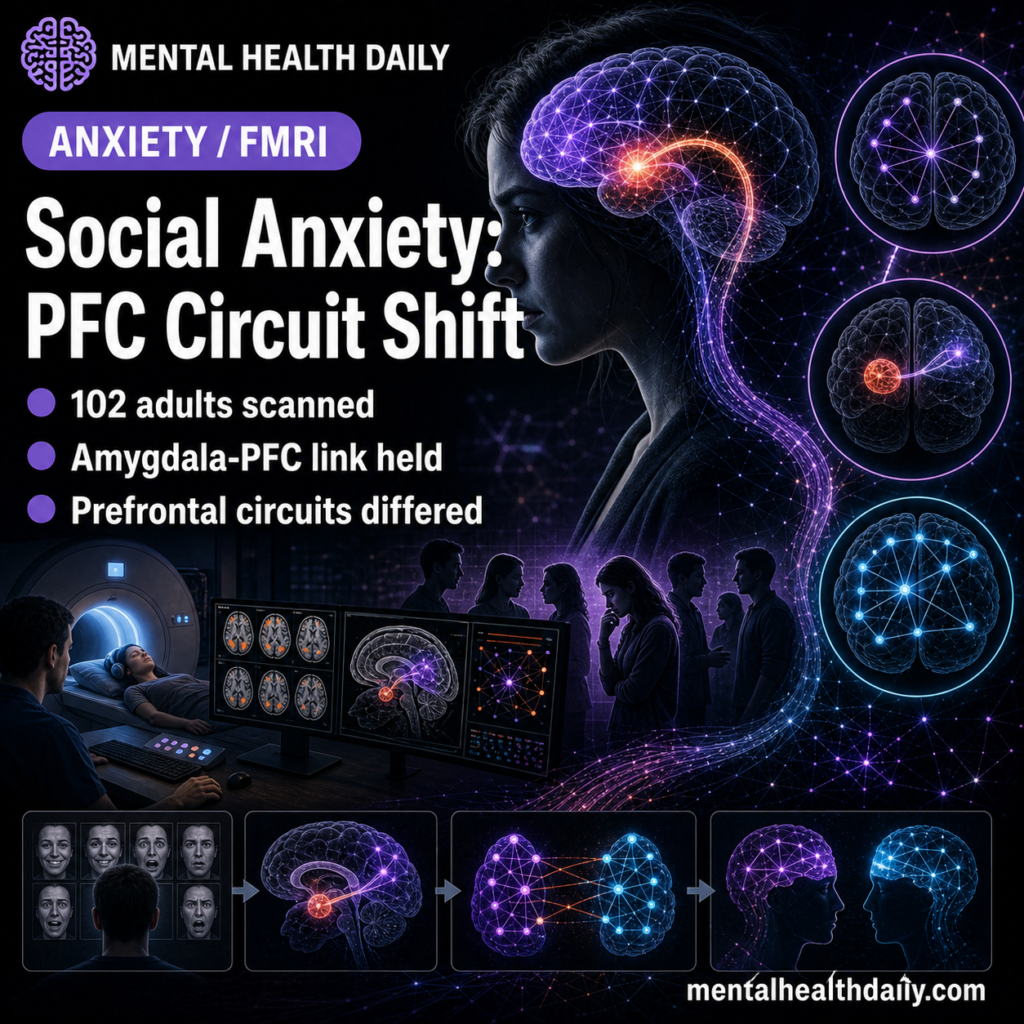
Specifically, the differences were:
- During observation of negative faces: Social anxiety patients exhibited stronger preSMA-to-dlPFC inhibition compared to controls. This suggests that even when merely observing emotional cues, the preSMA in SAD patients might be exerting a more pronounced suppressive influence on the dorsolateral prefrontal cortex, a region involved in working memory and cognitive control.
- During active emotion regulation: Two key differences emerged:
- Stronger preSMA-to-vmPFC excitatory coupling in SAD patients.
- Stronger vmPFC-to-preSMA excitatory coupling in SAD patients.
These findings indicate an increased and altered reciprocal excitatory interaction between the preSMA and vmPFC during attempts to regulate emotions. The vmPFC is critical for integrating emotion and cognition, while the preSMA plays a pivotal role in action selection and volitional control.
These intra-prefrontal changes suggest that while SAD patients successfully reduced negative ratings during regulation (with self-reported regulation success not differing significantly between groups), they achieved this through distinct neural pathways. The implication is not that "patients can’t regulate," but rather that "patients regulate via different prefrontal circuits," potentially reflecting a heightened and atypical engagement of cognitive control mechanisms.
The Role of the Pre-Supplementary Motor Area (preSMA)
The pre-supplementary motor area (preSMA), though traditionally associated with motor planning, has a well-established role in higher-order cognitive and affective control. It is involved in conflict monitoring (detecting when competing response options exist), response inhibition (suppressing unwanted actions), volitional behavioral adjustment, and preparing adaptive responses to salient stimuli.
The Schrammen findings can be interpreted as follows:
- The stronger preSMA-to-dlPFC inhibition during observation in SAD patients might reflect an initial, perhaps hyper-vigilant, attempt to inhibit or control cognitive processing even before active regulation is initiated. This could contribute to the characteristic overthinking and self-monitoring seen in social anxiety.
- The enhanced reciprocal excitatory coupling between preSMA and vmPFC during active regulation suggests a more effortful or altered strategy for integrating emotional information with behavioral control. This could imply that individuals with SAD engage these regions more intensely or differently to achieve the same regulatory outcome as healthy individuals, perhaps indicating a compensatory mechanism or a less efficient neural pathway.
Re-evaluating the Amygdala-Prefrontal Story
The Schrammen DCM result provides crucial context for the inconsistent findings from previous functional connectivity studies regarding amygdala-prefrontal connectivity in social anxiety. While some studies reported reduced vmPFC-amygdala connectivity in SAD, others found increased connectivity, and many reported no significant differences. These inconsistencies might, in part, be explained by the limitations of functional connectivity in determining directionality. What appeared as a "coupling" or "decoupling" might actually be complex, bidirectional influences that only DCM can disentangle.
The study does not claim the amygdala is unimportant in social anxiety. Indeed, meta-analyses consistently show amygdala hyperactivation to threatening faces in SAD. However, activation differences are distinct from connectivity differences. The amygdala can be more reactive without its coupling with the prefrontal cortex being fundamentally different in direction or strength. The Schrammen study specifically challenges the notion that amygdala-prefrontal connectivity is the key disrupted circuit, redirecting focus to the intricate dynamics within the prefrontal cortex.
Implications for Treatment and Research
While the immediate impact on clinical practice is subtle, the implications for future research and treatment development are profound. Current first-line treatments for social anxiety, such as cognitive-behavioral therapy (CBT) and selective serotonin reuptake inhibitors (SSRIs), remain the most evidence-supported interventions. However, this study offers a refined understanding of the neural targets upon which these treatments might be acting.
- Refining Treatment Mechanisms: CBT, with its emphasis on exposure and cognitive restructuring, aims to modify maladaptive thought patterns and behavioral responses. Understanding that intra-prefrontal circuits, particularly those involving the preSMA, are altered in SAD suggests that CBT might be effectively re-wiring these specific pathways to improve cognitive control and adaptive responses.
- Targeted Therapies: For future therapeutic development, this research opens the door to exploring interventions that specifically modulate prefrontal-prefrontal circuits. This could include novel pharmacotherapies designed to enhance or normalize connectivity within these regions, or advanced neuromodulation techniques (e.g., transcranial magnetic stimulation, TMS, or neurofeedback) that can precisely target the preSMA or its connections with other prefrontal areas.
- Patient Understanding: For patients who might have internalized a narrative of a "broken amygdala" or an uncontrollable threat response, this research offers a more complex and potentially empowering perspective. It emphasizes that the brain differences are distributed across multiple regions and involve a sophisticated interplay of control mechanisms, rather than a single, irretrievably "overactive" structure.
- Diagnostic Tools: Despite the statistical significance of the findings (pooled connectivity pattern predicted diagnostic group with r = 0.24, p = 0.009), the predictive accuracy is modest. This means that while the pattern carries valuable information for research, a brain scan is not yet a useful clinical tool for diagnosing social anxiety at the individual level. Clinical diagnosis continues to rely on DSM-5 criteria and validated scales like the Liebowitz Social Anxiety Scale.
Limitations and Future Directions
The Schrammen study, while highly impactful, has several limitations common to neuroimaging research:
- Single-site, single-task: Conducted at one university center using one emotion-regulation paradigm and a specific stimulus set (Karolinska Directed Emotional Faces). Generalizability across diverse populations, tasks, and stimuli requires further investigation.
- Self-chosen regulation strategy: While ecologically valid, allowing participants to choose their own strategy introduces variability, potentially diluting amygdala-prefrontal differences that might be specific to certain strategies like reappraisal.
- Mildly negative stimuli: The use of mildly negative faces might not fully engage amygdala-prefrontal pathways as robustly as stronger aversive stimuli (e.g., social rejection scenarios, public speaking imagery), which could potentially yield different group differences.
- Mixed medication status: A significant portion of SAD patients (18 of 61) were on antidepressants, while controls were medication-naive. This introduces potential confounding from chronic antidepressant use, which can alter brain connectivity.
- Simplified network model: The model included only five nodes. Other regions crucial for emotion regulation (e.g., insula, anterior cingulate cortex, hippocampus) were not modeled. The conclusion is that intra-prefrontal differences were more prominent within this specific 5-node network, not that amygdala-prefrontal connectivity is universally unimportant in social anxiety.
Future research will need to address these limitations through multi-site studies, diverse experimental paradigms, longitudinal designs to track treatment effects on these pathways, and more comprehensive network models incorporating additional brain regions.
Conclusion: A New Era in Understanding Social Anxiety
The 2026 Schrammen et al. study marks a significant milestone in social anxiety research. By employing Dynamic Causal Modeling, it has provided unprecedented insights into the directed flow of information within brain networks, fundamentally altering the understanding of SAD’s neural mechanisms. The revelation that intra-prefrontal circuits, particularly those involving the preSMA, are key differentiators between individuals with and without social anxiety, rather than direct amygdala-prefrontal dysregulation, redirects scientific inquiry.
This study underscores the brain’s remarkable complexity and the distributed nature of emotion regulation. While the amygdala remains an important player in threat detection, the nuanced control and regulation of social fear appear to be more intricately woven into the fabric of the prefrontal cortex itself. This new map of social anxiety promises to accelerate the development of more precise, mechanism-based treatments and fosters a deeper, more accurate understanding of this pervasive disorder.

