

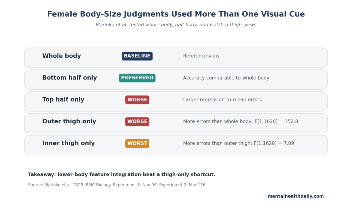
A recent study published in BMC Biology has significantly advanced our understanding of how individuals visually assess female body size, revealing that accurate judgments do not necessitate a full-body view but rely heavily on integrated information from the lower body. The two-experiment investigation, involving a total of 215 women, demonstrated that bottom-half views maintained accuracy comparable to whole-body views, while isolated views of inner or outer thighs introduced notable perceptual errors. This pivotal finding underscores the concept of lower-body feature integration, suggesting that the visual system combines multiple cues from the lower body rather than relying on a simplistic "thigh-only" shortcut, a distinction critical for refining body-image research and its clinical applications.
Understanding Body-Size Judgment and Its Clinical Significance
Body-size judgment refers to the cognitive process of estimating a body’s dimensions based on visual input. This perceptual aspect forms a crucial component of broader body-image research, which also encompasses phenomena like body dissatisfaction, compulsive body checking, avoidance behaviors, and anxiety-driven interpretations of one’s own physique. Disturbances in body image are clinically significant, particularly within the context of eating disorders such as anorexia nervosa and bulimia nervosa, where patients frequently misperceive or overestimate their body size. The underlying mechanism involves a complex interplay of visual perception, where the brain must discern which cues are relevant, assign appropriate weight to them, and manage the influence of recent visual experiences on subsequent judgments.
Historically, body-image disturbance has been recognized as a core feature of eating disorders. Pioneering work, such as Cash and Deagle’s meta-analysis, solidified its status as a measurable clinical feature across diverse eating-disorder populations, moving it beyond the realm of vague personality complaints. More contemporary research has shifted focus from generalized body dissatisfaction towards precise visual mapping and identifiable perceptual markers. Studies like that by Ralph-Nearman et al., which utilized digital body-image mapping in individuals with anorexia nervosa, have demonstrated a correlation between visual disturbance measures and illness severity, reinforcing the idea that body-image disturbance is quantifiable alongside self-reported experiences. The Marinko et al. study contributes to this evolving landscape by dissecting the specific visual cues involved in body-size perception.
Experiment 1: The Sufficiency of Lower-Body Information
The initial experiment in the BMC Biology study sought to determine whether a full-body view was essential for accurate size judgments or if partial views could suffice. Researchers, led by Marinko et al., employed a "bodyline task," a specialized visual-perception task where participants evaluated body images and categorized them along a size continuum. This task is particularly effective because it can distinguish between a person’s average judgment and more specific patterns of error.
After an initial screening process that excluded five participants who could not reliably differentiate body-size categories, the analysis proceeded with 99 women. These participants were presented with images under three viewing conditions: whole-body, top-half, and bottom-half. The images represented seven distinct body-size categories, ensuring a comprehensive range for assessment.
The results unequivocally showed that participants could clearly distinguish between different size categories, indicated by a highly significant statistical outcome (F(2.48, 242.83) = 2164.72, p < 0.001). While the main effect of view condition did not reach statistical significance independently (F(2, 196) = 2.74, p = 0.067), a crucial interaction between body size and view condition was observed (F(7.28, 713.32) = 37.99, p < 0.001). This interaction revealed a direct and practical pattern: judgments made from bottom-half-only views were remarkably accurate, comparable to those made from whole-body views. In stark contrast, top-half-only views led to substantially larger "regression-to-the-mean" errors, particularly noticeable at the extreme ends of the body-size spectrum (i.e., for very thin and very large bodies).
Regression-to-the-mean error, in this context, describes a phenomenon where extreme judgments tend to be pulled towards the average. When participants viewed only the top half of a body, their judgments for unusually thin or unusually large bodies became less precise, drifting closer to what they might perceive as an "average" body size. This suggests that the top half alone lacked critical visual information necessary to accurately differentiate extreme body sizes, unlike the lower half.
Experiment 2: The Insufficiency of Isolated Thigh Cues
Building on the findings of Experiment 1, the researchers posed a more granular question in their second experiment: if the lower half provides sufficient information for accurate judgments, can a single, isolated region within the lower body carry this information independently? To address this, 116 women participated in a comparison between whole-body viewing and isolated views of either the inner thigh or the outer thigh.
The results provided a clear answer: no, isolated thigh regions were insufficient. Both conditions involving isolated thigh regions — inner-thigh-only and outer-thigh-only — resulted in significantly worse body-size judgments compared to whole-body viewing. For regression-to-the-mean error, the outer-thigh comparison indicated a model with a shallower slope and greater error (F(1, 1620) = 152.8, p < 0.001). Similarly, the inner-thigh comparison also demonstrated poorer performance than whole-body viewing (F(1, 1620) = 262, p < 0.001).
Further analysis revealed a subtle but significant difference between the two isolated thigh views: inner-thigh-only viewing performed slightly worse than outer-thigh-only viewing in terms of regression-to-the-mean error (F(1, 1620) = 7.09, p = 0.008). Moreover, the phenomenon of "serial dependence bias" also broadened when only a single thigh region was visible. Serial dependence refers to the carryover effect where a current judgment is influenced by the previously viewed stimulus. In this study, the estimated serial-dependence amplitude (A) was 0.43 for whole-body viewing, increasing to 0.47 for outer-thigh viewing, and further to 0.64 for inner-thigh viewing. This indicates that judgments became more susceptible to the influence of the preceding image when visual information was restricted to isolated thigh regions, suggesting a higher degree of perceptual uncertainty.
The Power of Lower-Body Feature Integration
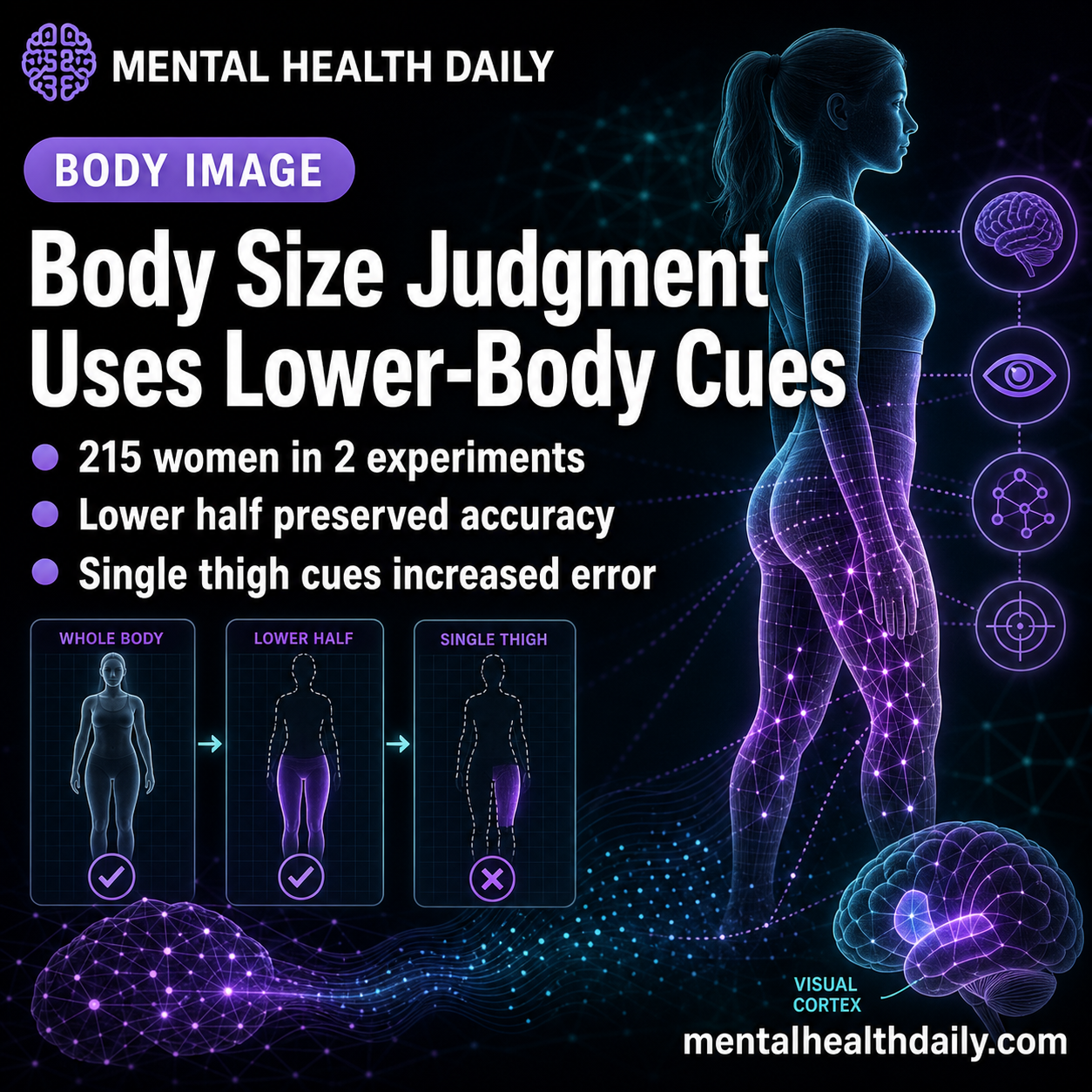
The central finding across both experiments points to the concept of "lower-body feature integration." This means that the human perceptual system appears to combine multiple visible cues originating from the lower body to form a comprehensive size estimate, rather than relying on a single anatomical patch. While a bottom-half view provides enough contextual information for accurate size estimation, stripping that view down to only one specific thigh region removes crucial contextual elements that the visual system typically utilizes. The integrity of the visual structure, including overall width, shape, and proportions provided by the entire lower half, appears essential for precise judgments. When this structure is fragmented, accuracy suffers.
Implications for Body-Image Research and Clinical Practice
The Marinko et al. study offers a refined understanding of the perceptual mechanisms underlying body-size judgment, with several important implications for both ongoing research and potential future clinical applications.
Not a Diagnostic Tool for Eating Disorders: It is crucial to emphasize that this study does not establish that lower-body cue weighting directly causes eating disorders. The participants in the study were not a clinical sample diagnosed with eating disorders, and the task involved judging standardized images of other female bodies, not a person’s own body under conditions of anxiety, mirror exposure, social comparison, or restrictive eating pressure. Therefore, the study does not offer a direct clinical test or diagnostic criterion for body-image disturbance.
Refining Methodological Approaches: The primary contribution of this research is methodological. It challenges the assumption that broad body dissatisfaction or a simple global body outline sufficiently captures the entire spectrum of perceptual problems in body image. Instead, it suggests that body-image measures should explicitly investigate which specific visual cues individuals utilize and how these cues are integrated. The weighting of lower-body cues, as identified in this study, could represent one measurable component within a larger, more complex perceptual system.
Alignment with Prior Visual-Cue Studies: This study aligns well with previous research emphasizing feature integration in visual perception. For instance, Irvine et al. utilized eye-tracking and visual masking to explore which cues drive self-assessment of body size. Their work highlighted that an observer’s gaze fixation point is not necessarily synonymous with the information the visual system actually processes. They found that judgment accuracy often depends on body-edge cues that define width and shape, even if observers look centrally. A separate 2024 study by Marinko et al. further tested whether internal body features contribute useful information beyond the silhouette shape. That research concluded that removing internal features increased body-size judgment errors and altered serial-dependence patterns, arguing against a simplistic "outline-only" account. The current 2025 lower-body study adds another specific piece to this puzzle, demonstrating that full lower-body information can maintain accuracy, whereas isolated fragments cannot. Collectively, this body of evidence strongly supports a feature-integration model, wherein body-size perception relies on multiple visual cues, and accuracy diminishes when too much of that essential cue structure is removed.
Countering Misinterpretations: Beyond "Thigh Obsession"
A common pitfall in disseminating scientific findings to the public is oversimplification. This paper could easily be misconstrued as suggesting that people judge female body size predominantly by looking at thighs. The actual results, however, are far more nuanced and cautious. While Experiment 1 demonstrated that bottom-half information was sufficient for accurate judgment, Experiment 2 definitively showed that a single, isolated thigh region was insufficient.
This crucial distinction fundamentally alters the interpretation:
- Evidence Strength: This is a rigorously controlled visual-perception study, adept at identifying task-specific judgment mechanisms. It is not designed to diagnose body-image disturbance, establish causality in eating disorders, or dictate specific body regions for clinical treatment.
- Clinical Caution: Translating these findings into tangible eating-disorder care requires further dedicated studies that directly investigate self-body perception, its relationship to symptom severity, and its responsiveness to treatment. Judging standardized images of others is distinct from the intensely personal and often emotionally charged experience of judging one’s own body.
The practical takeaway for the scientific community is therefore methodological: body-image research should prioritize measuring the specific visual cues individuals employ and how these cues are integrated, rather than making broad assumptions about the equivalence of global dissatisfaction, gaze patterns, and perceptual accuracy. The demonstrated weighting of lower-body cues represents a measurable component of this intricate perceptual system.
Future Directions and Unanswered Questions
This study opens several avenues for future research. One critical next step involves replicating these findings in clinical populations, such as individuals diagnosed with anorexia nervosa, bulimia nervosa, or body dysmorphic disorder. It remains to be seen whether these populations exhibit different patterns of cue weighting or a heightened sensitivity to specific lower-body features. Researchers might also explore gender differences in body-size judgment, as this study focused exclusively on female participants judging female bodies.
Furthermore, these findings could inform the development of more sophisticated and targeted assessment tools for body image. Instead of relying solely on self-report questionnaires, future assessments might incorporate tasks that probe the integration of visual cues, providing a more objective measure of perceptual disturbance. This could, in turn, lead to more individualized therapeutic interventions that address specific perceptual biases, potentially enhancing the effectiveness of treatments for eating disorders and other body-image concerns.
The study also prompts a deeper exploration into why bottom-half views prove more effective than top-half views. The most plausible explanation is that the lower body preserves a greater amount of critical visual information related to overall width and shape, which participants utilize to infer body size. Top-half views, conversely, lose enough of this relevant information that judgments for very thin or very large bodies are disproportionately pulled towards an average-size perception.
In conclusion, the research by Marinko et al. significantly sharpens the perceptual dimension of body-image research. It demonstrates that accurate female body-size judgment is neither purely holistic nor reducible to a single body part. Instead, accuracy is profoundly dependent on the integrated processing of multiple visual cues from the lower body, offering a nuanced and important contribution to a field with profound clinical implications.

