

A groundbreaking preprint from the Scottish health system has unveiled a sophisticated application of natural language processing (NLP) to significantly enhance stroke subtyping from routine medical imaging reports, leading to crucial insights into post-stroke dementia risk, particularly for lobar intracerebral hemorrhage (ICH). This innovative methodology, detailed in a 2026 study by Hosking et al., analyzed vast datasets of CT and MRI reports, revealing that lobar ICH carries a substantially higher risk of dementia beyond six months post-event, with an adjusted hazard ratio (aHR) of 3.49 compared to carefully matched controls. Crucially, the NLP-driven approach dramatically reduced the rate of unspecified stroke coding from 26.1% to a mere 3.4%, paving the way for more precise prognostication and highly individualized patient care pathways across Scotland’s health system.
Unlocking Precision in Stroke Classification with Natural Language Processing
The core innovation of the Scottish study lies in its deployment of natural language processing (NLP) software to meticulously analyze the free-text narratives embedded within radiology reports. Unlike traditional administrative coding systems that often categorize stroke broadly for billing and administrative purposes, radiologists’ reports contain rich, granular detail regarding the precise anatomical location and specific characteristics of a stroke event. NLP, in this context, functions as a sophisticated computational tool designed to read and extract structured, actionable information from unstructured clinical text. In the Hosking et al. study, this meant parsing thousands of CT and MRI head scan reports to assign specific stroke subtypes, moving beyond generic labels to identify categories such as deep ischemic stroke, cortical ischemic stroke, deep intracerebral hemorrhage, and lobar intracerebral hemorrhage.
This level of detailed subtyping is far from a mere cosmetic improvement in data management. It fundamentally alters the ability of healthcare systems and researchers to derive accurate prognoses from routine clinical data. By transforming the qualitative, descriptive language of radiology into quantifiable, structured data points, the NLP system empowers clinicians and researchers to identify subtle yet significant differences in patient outcomes that were previously obscured by broad, undifferentiated classifications. The dramatic reduction in unspecified stroke coding—from over a quarter of all cases to less than 4%—represents a monumental leap in data utility, offering an unprecedented level of clarity for understanding the varied trajectories of stroke recovery and potential long-term complications. This improved data fidelity can inform better resource allocation, targeted interventions, and enhanced patient counseling.
The Scottish Cohort: A Foundation for Large-Scale Insights
The research leveraged an extensive and robust dataset drawn from the Scottish national health system, linking head-scan reports with a comprehensive array of other crucial health records. This integrated approach allowed for a holistic view of patient health trajectories following a stroke, encompassing data on hospital readmissions, prescription histories, death records, and cancer registry information. The source population for the study was immense, encompassing 785,331 individuals who had undergone head scans, with 64,219 of these individuals subsequently identified as having clinical stroke phenotypes.
The demographic profile of the stroke cohort reflected a typical aging population susceptible to stroke, with a mean age of 73.4 years and a near-equal gender distribution, with 49.5% being male. Such a large and diverse population base lends significant statistical power and generalizability to the study’s findings, moving beyond small-scale observational studies to establish robust population-level trends. Through the sophisticated application of the NLP system, the researchers successfully subtyped a significant number of stroke events, including 12,616 deep ischemic strokes, 14,103 cortical ischemic strokes, 1,814 deep intracerebral hemorrhages, and 1,456 lobar intracerebral hemorrhages. These precisely classified categories enabled the researchers to perform a granular analysis of subsequent health outcomes, revealing critical distinctions in risk profiles across different stroke types.
Lobar Intracerebral Hemorrhage: A Clear Signal for Elevated Dementia Risk
One of the most striking and clinically significant findings of the study was the pronounced link between lobar intracerebral hemorrhage (ICH) and a substantially elevated risk of developing dementia. Beyond the initial six-month acute recovery period, individuals who experienced a lobar ICH demonstrated an adjusted hazard ratio (aHR) of 3.49 for developing dementia, with a 95% confidence interval (CI) of 2.30-5.29, when compared against carefully matched control groups. An adjusted hazard ratio is a statistical measure that accounts for other measured differences between groups, providing a more accurate comparison of the likelihood of an event (in this case, dementia) occurring over time. This statistically significant association underscores the unique vulnerability of patients with lobar ICH to long-term cognitive decline.
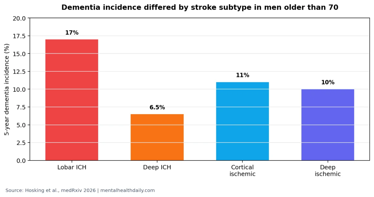
To translate these statistical figures into more tangible terms for clinicians and patients, the study also provided absolute risk contrasts, offering valuable insight into the practical implications. For men older than 70, the 5-year cumulative incidence of dementia varied significantly across different stroke subtypes:
- Lobar ICH: A high 17% incidence.
- Cortical Ischemic Stroke: An 11% incidence.
- Deep Ischemic Stroke: A 10% incidence.
- Deep ICH: A comparatively lower 6.5% incidence.
These figures clearly illustrate that individuals experiencing lobar ICH face nearly double the dementia risk compared to those with cortical or deep ischemic strokes, and almost triple the risk compared to deep ICH. This stark difference in absolute risk provides compelling evidence for the necessity of targeted monitoring and personalized intervention strategies following a stroke.
Understanding the Biological Basis: Why Location Matters
The distinction between lobar and deep intracerebral hemorrhage is not merely an anatomical detail; it reflects fundamentally different underlying pathologies, which in turn significantly influence the risk of subsequent complications like dementia. Intracerebral hemorrhage refers to bleeding directly into the brain tissue. Lobar hemorrhage occurs in the outer regions of the brain’s lobes, typically closer to the cerebral cortex and its associated networks. In contrast, deep hemorrhage affects deeper brain structures such as the basal ganglia, thalamus, or brainstem.
This distinction in location is critical because lobar hemorrhages are often associated with conditions like cerebral amyloid angiopathy (CAA) and cortical network injury. Cerebral amyloid angiopathy is a progressive condition characterized by the abnormal accumulation of amyloid proteins in the walls of small and medium-sized blood vessels within the brain’s cortex and leptomeninges, making them fragile and prone to bleeding. CAA is a recognized risk factor for recurrent lobar hemorrhages, cerebral microbleeds, cortical superficial siderosis, and, importantly, is strongly implicated in cognitive decline and dementia. While the Hosking analysis did not directly diagnose CAA in individual patients, the strong and specific association between lobar ICH and dementia risk is biologically plausible given the known link between CAA, lobar hemorrhages, and neurodegenerative processes.
Deep hemorrhages, on the other hand, are more frequently linked to chronic hypertensive small-vessel disease, where long-standing high blood pressure damages the tiny arteries and arterioles in the deeper brain structures. These differing vascular substrates lead to distinct patterns of brain injury and, consequently, different long-term sequelae, including significant variations in dementia risk. The study’s ability to precisely differentiate these subtypes at a population scale validates the critical clinical and pathological significance of these distinctions, moving towards a more nuanced understanding of stroke’s impact.
Beyond Dementia: Stroke Subtype Influences Epilepsy and Myocardial Infarction Risk
The implications of precise stroke subtyping extend beyond the significant findings regarding dementia, revealing varied risks for other critical post-stroke complications, including epilepsy and myocardial infarction (MI). The study found significant differences in the incidence of these outcomes across stroke subtypes, further underscoring the necessity of individualized follow-up care.
- Epilepsy Risk: Early epilepsy after lobar ICH was remarkably higher, with an aHR of 10.98 (95% CI 6.82-19.08), presenting a significantly greater risk compared to an aHR of 3.42 (95% CI 1.49-7.86) after deep ICH. This dramatic difference in epilepsy risk highlights the acute need for specific counseling and proactive monitoring for patients with lobar ICH, given their cortical involvement.
- Myocardial Infarction Risk: Patients experiencing a cortical ischemic stroke showed a significantly increased risk of early myocardial infarction, with an aHR of 4.6 (95% CI 3.35-6.31). This finding suggests a need for heightened cardiovascular evaluation and aggressive preventive management in this specific subgroup of stroke survivors, reflecting the complex interplay between cerebrovascular and cardiovascular health.
- Recurrent Stroke: The risk of readmission due to a recurrent stroke was also notably higher after lobar ICH compared to deep ICH, with an aHR of 1.71 (95% CI 1.15-2.54). This further emphasizes the unique and complex risk profile of lobar ICH patients, extending to the likelihood of repeat cerebrovascular events.
These findings collectively argue for a more granular, subtype-aware approach to stroke follow-up. The specific type of stroke an individual experiences can and should inform the intensity and focus of cognitive monitoring, seizure counseling, recurrent-stroke surveillance, comprehensive cardiovascular evaluation, and the prioritization of dementia-risk communication. This precision medicine approach moves beyond a "one-size-fits-all" model for stroke survivors.
The Role of NLP: Augmenting, Not Replacing, Clinical Judgment
It is crucial to contextualize the nature of the NLP methodology employed in this study. This research, presented as a preprint (meaning it has yet to undergo full peer review), utilized rules-based NLP applied to existing radiology-report text. The system did not directly interpret raw CT or MRI images; rather, it efficiently extracted and categorized the diagnostic information that radiologists had already meticulously documented in their reports. This distinction is vital: the NLP system functions as an incredibly efficient and consistent data extractor, transforming unstructured clinical narratives into structured, analyzable data points.
While profoundly powerful for population-scale research and improving audit coding, this approach inherently inherits the quality, completeness, and local conventions of the original radiology reports. Factors such as variations in local language, the availability and quality of scans, and the completeness of outpatient dementia diagnoses can influence the extracted data. Therefore, the NLP system is best understood as a sophisticated tool for enhancing health-system research and improving data utility, rather than a replacement for individualized clinical assessment. Patients still require comprehensive clinical evaluation, thorough imaging review by a radiologist, detailed cognitive history taking, and personalized follow-up care. The NLP-derived subtype data adds a layer of precision to the risk stratification map, but ultimately, clinical assessment remains paramount for patient-level interpretation and informed decision-making.
Bridging the Gap: Why Routine Records Often Miss Critical Prognostic Details
A persistent and pervasive challenge in modern healthcare data management is the disparity between the rich, nuanced detail captured in clinical narratives, such as radiology reports, and the often-simplified, broad categories used for administrative, billing, and public health reporting purposes. Administrative diagnosis codes typically flatten complex medical conditions into generic categories like "stroke," "ischemic stroke," or "intracerebral hemorrhage." In stark contrast, a radiologist’s report might specify whether a lesion was cortical, deep, lobar, lacunar, hemorrhagic, or recurrent – details that are profoundly important for understanding prognosis and guiding individualized treatment strategies.
This fundamental mismatch creates significant problems for both epidemiological research and the quality of patient care. If a substantial proportion of strokes (e.g., the original 26.1% unspecified rate in this study) remain undifferentiated in structured electronic health record fields, a health system loses the ability to easily compare dementia risk by precise subtype, target appropriate follow-up care, or even identify which patients require specific counseling, such as for seizure prevention. By dramatically reducing unspecified stroke coding to a mere 3.4%, the NLP method renders the same routine record system exponentially more clinically informative and actionable, unlocking a wealth of previously inaccessible data for clinical decision-making and public health planning.
The method, while highly effective in its practical application, might be considered "less glamorous" than some of the "AI in medicine" headlines imply. It is not about developing artificial general intelligence mimicking human thought, but rather about rules-based text extraction solving a very concrete, widespread, and costly documentation problem. Radiologists already possess and meticulously record these crucial subtype clues; the software simply makes these clues systematically usable at a population scale, unlocking their latent value for patient care, quality improvement, and public health research.
Transforming Post-Stroke Follow-Up: A Subtype-Aware Approach to Care
The findings from this Scottish preprint strongly advocate for a significant paradigm shift in post-stroke follow-up care, moving away from a uniform, generic approach to one that is finely tuned to the specific stroke subtype. The distinct profiles observed for dementia, epilepsy, readmission, and myocardial infarction across lobar ICH, cortical ischemic stroke, deep ICH, and deep ischemic stroke necessitate differentiated and personalized care pathways.
For patients who have experienced a lobar intracerebral hemorrhage, particularly older adults, the cognitive signal for dementia is unequivocally the strongest. A 17% 5-year dementia incidence in men older than 70 is a significant enough risk to warrant proactive and comprehensive measures. This should translate into scheduled, regular cognitive screening, comprehensive family education regarding the early signs of cognitive changes, a thorough medication review, aggressive management of all modifiable vascular risk factors, and a low threshold for referral to a specialized memory clinic should any cognitive symptoms emerge. The overarching goal is early detection and intervention, rather than waiting for profound cognitive decline to become evident years later, when interventions may be less effective.
Similarly, for individuals with a cortical ischemic stroke, the elevated risk of myocardial infarction and recurrent vascular events suggests that intensive cardiovascular evaluation and aggressive preventive strategies should be given particular prominence alongside cognitive monitoring. For lobar ICH patients, specialized seizure counseling becomes especially relevant given the significantly higher risk of early epilepsy compared to deep ICH, requiring proactive patient education and potential anticonvulsant management. The stroke subtype label, while not a complete care plan in itself, serves as a powerful and essential guide for prioritizing which risks require immediate attention and focused discussion with the patient and their caregivers.
The operational lesson derived from this research is clear and impactful: structured health records must evolve to consistently preserve critical anatomical detail. When crucial information regarding stroke location is lost within generic coding, the ability to accurately predict and effectively manage patient prognosis is severely hampered. Conversely, when the detailed information from radiology reports is systematically recovered and utilized through technologies like NLP, patients can be accurately stratified into clinically meaningful risk categories, leading to more precise, effective, and ultimately, more humane care.
It is essential to interpret the dementia finding as a tool for risk stratification rather than an unchangeable fate. Numerous factors contribute to cognitive outcomes after stroke, including older age, pre-stroke cognitive status, the occurrence of recurrent strokes, overall lesion burden, pre-existing vascular risk factors, educational attainment, episodes of delirium, depression, specific medications, and access to rehabilitation services. While stroke subtype significantly improves the "risk map," patient-level history and ongoing comprehensive

