

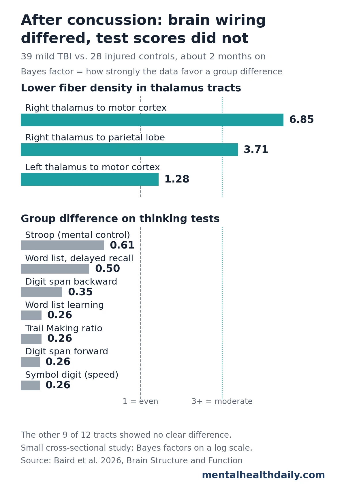
New research highlights that adults recovering from a concussion may still exhibit subtle alterations in brain wiring two months post-injury, specifically in white matter tracts connecting the thalamus to the brain’s cortex, despite achieving normal scores on standard cognitive tests. This groundbreaking finding, derived from a 2026 study comparing individuals with mild traumatic brain injury (mTBI) to a control group who experienced accidents without head trauma, underscores the "invisible" nature of concussions and challenges the assumption that normal cognitive function equates to complete structural recovery.
Understanding the Enigma of Concussion
Concussion, a form of mild traumatic brain injury (mTBI), is a complex neurophysiological disturbance resulting from a direct blow to the head or an indirect force causing rapid acceleration and deceleration of the brain. Often referred to as an "invisible injury," concussions frequently present without visible damage on conventional imaging techniques like CT scans or standard MRI. Symptoms can range from headaches and dizziness to difficulties with concentration and memory, though these typically resolve within a few weeks to months for the majority of individuals. However, a subset of patients experiences persistent symptoms, raising questions about the underlying biological mechanisms and the long-term implications of these injuries.
Globally, concussions are a significant public health concern, affecting millions annually. Sports-related concussions, military blast injuries, and falls are common causes. The challenge in managing concussions lies in their heterogeneous presentation and the difficulty in objectively assessing the injury’s severity and recovery trajectory. The absence of clear biomarkers or structural damage on routine scans has historically made it difficult to provide definitive answers to patients regarding the state of their brain post-injury, particularly when cognitive symptoms have seemingly resolved.
The Thalamus: A Central Hub Under Scrutiny
At the heart of this new research lies the thalamus, a pair of egg-shaped structures nestled deep within the brain. Functioning as a critical relay hub, the thalamus processes and transmits sensory and motor signals to and from nearly every part of the cerebral cortex. Its vital roles extend to attention, motor control, and processing speed, making it indispensable for complex cognitive functions.
The nerve fiber bundles connecting the thalamus to the cortex are known as thalamocortical tracts. These white matter pathways are particularly vulnerable to the shearing and twisting forces that occur during a head injury. Due to the thalamus’s central location, these long tracts can be subjected to significant mechanical stress during a concussive event, potentially leading to subtle but impactful structural damage. Previous studies have increasingly pointed to the thalamus as a key region affected by concussion, with research noting changes in thalamic volume, connectivity, and metabolic activity. This growing body of evidence emphasizes the importance of a deeper understanding of how concussions impact this critical brain structure and its extensive network of connections.
Advanced Imaging Techniques Unveil Hidden Damage
Traditional neuroimaging, such as CT scans and conventional MRI, often fails to detect the microscopic damage characteristic of concussions. These methods are adept at identifying gross structural abnormalities like hemorrhages or contusions but lack the resolution to visualize subtle alterations in the brain’s intricate white matter pathways. This limitation has historically hindered a comprehensive understanding of concussion pathophysiology.
To overcome these challenges, researchers have turned to advanced neuroimaging techniques, most notably diffusion MRI (dMRI). Diffusion MRI works by tracking the movement of water molecules, which tend to diffuse more freely along the direction of nerve fibers (axons) than across them. By mapping this anisotropic water movement, dMRI can infer the orientation, integrity, and density of white matter tracts.
However, even older dMRI methods had limitations. They often averaged fiber directions within each small volumetric unit of brain tissue, known as a voxel. This averaging could blur the picture, particularly in areas where multiple fiber bundles intersect or cross, leading to an underestimation of subtle damage.
This is where Fixel-Based Analysis (FBA) represents a significant leap forward. FBA is a cutting-edge diffusion MRI technique that refines the analysis by splitting each voxel into its constituent fiber bundles, called "fixels." Each fixel can then be measured independently, allowing for unprecedented precision in assessing individual fiber bundles, even in complex crossing fiber regions. The primary measure employed in this study was fiber density (specifically, fiber density and cross-section, or FOD-FDC), which quantifies the structural integrity and packing density of nerve fibers within these specific bundles. By using FBA, researchers can detect more subtle changes in white matter integrity that would be missed by conventional dMRI approaches, providing a more granular view of the brain’s microstructural architecture after injury.
The Melbourne Concussion Study: A Rigorous Approach
The study in question, led by Maggie Baird et al. and conducted in Melbourne, Australia, employed a robust methodology designed to isolate the effects of head injury. Between 2017 and 2023, researchers recruited adult participants admitted to two Melbourne hospitals following an injury. The study compared two distinct groups: 39 individuals diagnosed with mild traumatic brain injury (concussion) and 28 control participants who had sustained injuries from accidents but without any head trauma.
A crucial strength of this study design was the inclusion of injured controls. This approach is vital because general trauma, even without head injury, can lead to symptoms such as pain, fatigue, and psychological distress, all of which can influence cognitive test performance. By comparing the concussion group to controls who experienced similar levels of physical injury and potential distress, the researchers were better able to isolate the specific neurological effects attributable to the head injury itself, rather than confounding factors.
Participants underwent advanced brain imaging using Fixel-Based Analysis approximately two months after their respective injuries. This specific timeline was chosen to capture a phase where initial acute symptoms might have subsided, but potential longer-term structural changes could still be present. The detailed imaging and cognitive assessments allowed for a comprehensive evaluation of both structural integrity and functional performance.
Revealing Altered Fiber Density in Thalamic Tracts
The core findings of the Melbourne study revealed specific and localized white matter changes in the concussion group. After adjusting for age and sex, individuals who had experienced a concussion showed lower fiber density in two distinct right-sided thalamocortical tracts: the pathway connecting the thalamus to the motor cortex and the pathway connecting the thalamus to the parietal lobe.
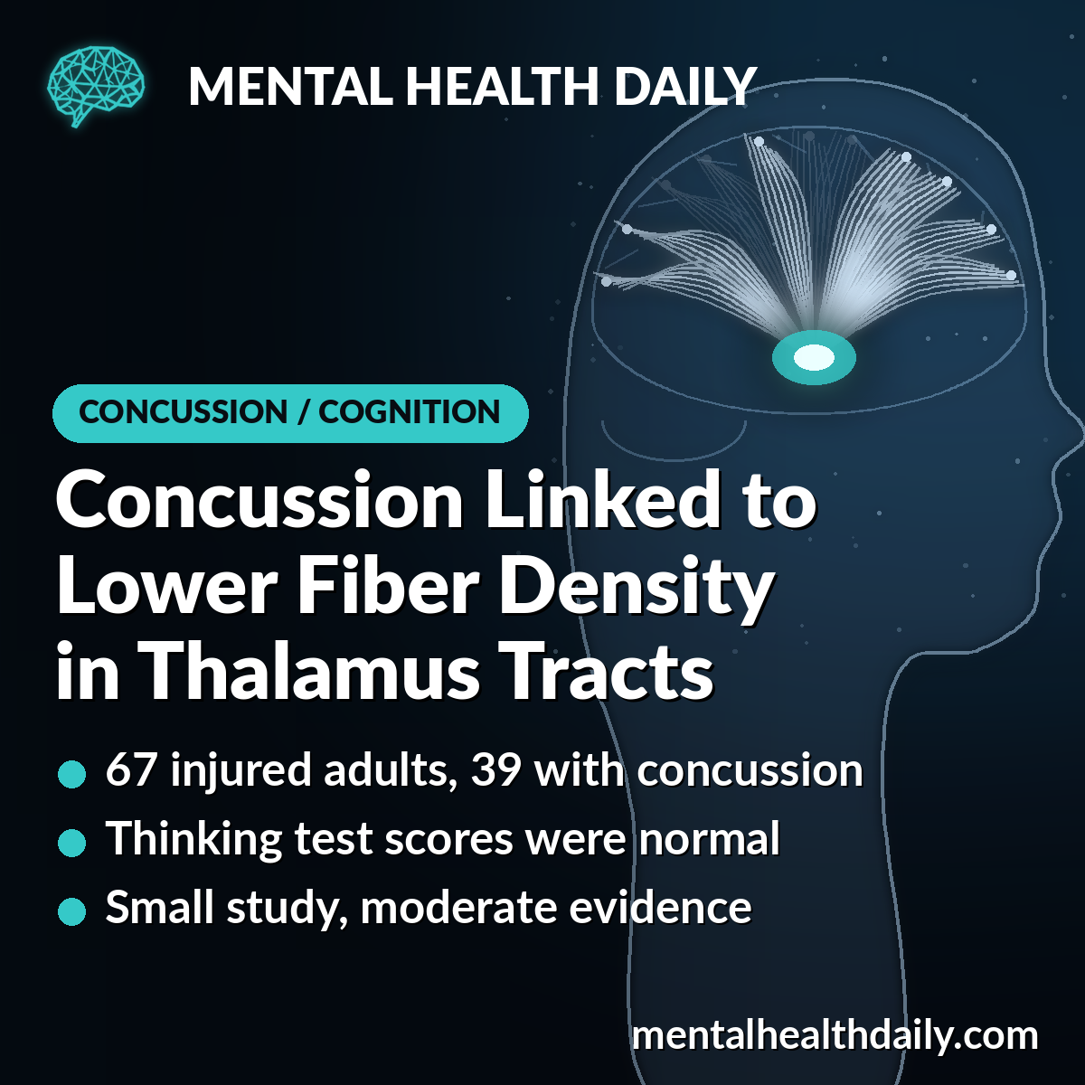
To quantify the evidence for these differences, the researchers utilized Bayes factors. A Bayes factor compares the likelihood of the data fitting a model where "the groups differ" versus a model where "the groups do not differ." A value above 3 is typically considered moderate evidence for a difference, while a value below 1/3 suggests evidence for no difference. In this study, the Bayes factor for lower fiber density in the right thalamus-to-motor-cortex tract was 6.85, and for the right thalamus-to-parietal tract, it was 3.71, both indicating moderate evidence for a significant reduction in fiber density in the concussion group. The motor cortex is crucial for planning, controlling, and executing voluntary movements, while the parietal lobe plays a key role in integrating sensory information, spatial awareness, and directing attention.
Interestingly, the other nine thalamocortical tracts examined did not show a clear difference between the groups. Furthermore, a whole-brain analysis, which tested every fixel across the entire brain, did not yield any significant differences after correcting for multiple comparisons. This suggests that the impact of concussion might manifest as subtle, diffuse changes spread along entire tracts, which become apparent when averaged over specific pathways but are too faint to be detected as focal points of damage.
The researchers proposed a compelling timeline for these fiber density changes: an initial increase in density in the immediate days post-concussion due to swelling, followed by a decrease if axonal degeneration occurs. The observed lower fiber density at the two-month mark aligns with this hypothesized later, lower phase, indicating a potential degenerative process in the white matter pathways.
Normal Cognitive Scores: A Deceptive Outcome
Perhaps one of the most intriguing and clinically significant findings of the study was the complete absence of detectable cognitive deficits in the concussion group. Both the mTBI participants and the control group completed a battery of seven measures drawn from five standard neuropsychological tests. These tests assessed various cognitive domains, including attention (digit span), processing speed (symbol digit), verbal learning and recall, mental control (Stroop), and task switching (Trail Making).
Across all these measures, there was no statistically significant difference between the concussion and control groups. The Bayes factors for group differences on these cognitive tests ranged from 0.26 to 0.61, all falling within the range favoring no difference or being inconclusive. For instance, the concussion group averaged 66.3 on the symbol digit speed test, while the control group averaged 65.3, demonstrating virtually identical performance.
This outcome is consistent with the typical recovery trajectory for most individuals after a concussion, where cognitive performance generally returns to baseline within approximately three months. However, the juxtaposition of normal cognitive scores with demonstrable structural changes in white matter tracts presents a critical paradox. It suggests that functional recovery, at least as measured by standard neuropsychological tests, does not necessarily equate to a complete restoration of underlying brain structure. The brain may be compensating for subtle damage, or the existing tests may not be sensitive enough to detect functional consequences of such nuanced structural alterations at this specific time point.
The study also delved into whether fiber density correlated with test scores in the same way for both groups. It found that these links did not always align, with some "exploratory" results suggesting altered brain-test relationships in the concussion group. However, the researchers noted that the expected pattern in controls (higher fiber density, better scores) often did not appear, possibly due to the confounding effects of pain, fatigue, and distress from their own injuries. This highlights the complexity of correlating brain structure with cognitive function, especially in populations experiencing generalized trauma.
The Thalamus: A Recurring Theme in Concussion Research
The findings from the Melbourne Concussion Study reinforce a growing consensus in the scientific community: the thalamus is a particularly vulnerable and crucial area in concussion research. Other studies have consistently highlighted its involvement:
- Thalamic Volume Loss: Some research has shown that individuals with a history of concussion can exhibit subtle reductions in thalamic volume, suggesting a long-term impact on the size and health of this structure.
- Altered Connectivity: Advanced functional MRI studies have revealed changes in the functional connectivity between the thalamus and other brain regions post-concussion, indicating disruptions in how different parts of the brain communicate.
- Metabolic Changes: Magnetic resonance spectroscopy (MRS) studies have detected alterations in metabolites within the thalamus, reflecting changes in brain chemistry and cellular health after mTBI.
- Role in Post-Concussion Syndrome: The thalamus’s involvement in sensory processing, pain modulation, and attention makes it a plausible candidate for contributing to persistent post-concussion symptoms such as chronic headaches, light sensitivity, and difficulty concentrating.
Collectively, these studies firmly establish the thalamus as one of the brain areas most profoundly affected by concussion. However, the precise relationship between these structural and functional changes and the manifestation of symptoms, as well as the long-term recovery trajectory, remains an active area of investigation.
Implications for Diagnosis, Treatment, and Future Research
The Melbourne Concussion Study carries significant implications for our understanding and management of mTBI. The revelation that normal cognitive test scores two months post-concussion do not guarantee a structurally pristine brain challenges existing paradigms and opens new avenues for clinical practice and research.
Clinical Significance: For individuals recovering from a concussion, these findings underscore that the absence of overt cognitive deficits does not necessarily mean the brain has fully healed. This knowledge can help validate the experiences of patients who report lingering subjective symptoms despite seemingly normal objective test results. Medical professionals must be aware that subtle wiring changes may still be present, and the link between brain structure and test performance might be altered, even if standard assessments appear reassuring.
Diagnostic Potential: While Fixel-Based Analysis is not yet a clinical diagnostic tool, this research highlights its potential as a highly sensitive biomarker for detecting subtle white matter damage in concussion. In the future, such advanced imaging techniques could potentially aid in more objective diagnosis, risk stratification, and personalized management plans for patients, especially those at risk for prolonged recovery or cumulative injury effects.
Understanding Recovery: The study contributes to a more nuanced understanding of concussion recovery, suggesting it is a multi-faceted process involving both functional adaptation and underlying structural changes. It emphasizes the need for longitudinal studies to track these subtle structural changes over extended periods and correlate them with long-term functional outcomes, including the development of post-concussion syndrome or increased vulnerability to future brain injuries.
Therapeutic Avenues: A deeper understanding of specific tract damage could eventually lead to the development of targeted therapies or rehabilitation strategies. If specific pathways are consistently affected, interventions aimed at promoting neural plasticity or repairing white matter could be explored.
Limitations and Future Directions: The researchers acknowledge several limitations, including the moderate sample size, the focus on a single time point (two months post-injury), and the broad range of injury severity within the mTBI group. The study also did not explicitly correlate the observed structural changes with specific symptoms, leaving the question of their direct clinical relevance open. Future research will need to address these limitations by conducting larger, multi-center studies, incorporating longitudinal follow-up at various time points post-injury, and more rigorously linking structural changes to clinical symptoms and functional outcomes. Investigating how these changes might evolve over months or even years will be crucial for understanding the long-term prognosis of concussions.
In conclusion, the Melbourne Concussion Study offers compelling evidence that even after seemingly complete cognitive recovery, subtle structural alterations in critical brain pathways can persist following a concussion. This research serves as a powerful reminder of the brain’s complex vulnerability and resilience. While Fixel-Based Analysis is a research tool, not a clinical test, its findings emphasize the importance of continued medical vigilance for anyone experiencing lingering symptoms such as headaches, fatigue, concentration difficulties, or slowed thinking after a concussion. These subtle changes underscore the ongoing need for comprehensive care and advanced research to fully unravel the long-term impact of this pervasive "invisible injury."

