

A groundbreaking dataset, encompassing recordings from an astonishing 33,028 neurons across 40 mice, has revealed that the neural circuits responsible for tracking a remembered smell are not confined to a single brain region but are instead distributed throughout the entire mouse brain. This monumental collection of neural activity, made publicly available in a 2026 data paper, offers an unprecedented resource for neuroscientists worldwide to delve into the intricate mechanisms of working memory, a cognitive function fundamental to daily life and frequently impaired in neurological and psychiatric conditions. The release of this comprehensive dataset marks a significant stride in the burgeoning field of open science, democratizing access to high-fidelity neural recordings and accelerating collaborative research into one of the brain’s most elusive processes.
Understanding Working Memory: The Brain’s Ephemeral Scratchpad
Working memory, often described as the brain’s short-term "scratchpad," is the cognitive system responsible for temporarily holding and manipulating information for a few seconds so that it can be used for ongoing tasks. Whether it’s remembering a phone number just long enough to dial it, following a complex set of instructions, or mentally calculating a tip, working memory is continuously engaged. Its efficiency is paramount for higher-order cognitive functions such as reasoning, problem-solving, and decision-making. Consequently, impairments in working memory are a hallmark symptom of numerous conditions, including schizophrenia, attention-deficit/hyperactivity disorder (ADHD), depression, and early-stage Alzheimer’s disease, making its underlying neural mechanisms a critical area of investigation for mental health research.
The scientific journey to understand working memory dates back decades. Early theories often posited that specific, localized brain regions were solely responsible for storing and processing short-term information. Pioneering work in the 1970s, particularly by Joaquín Fuster and George Alexander in 1971, began to challenge this localized view. Their seminal research, involving recordings from the prefrontal cortex and thalamus of monkeys performing memory tasks, demonstrated that certain neurons exhibited sustained firing activity during memory delays, maintaining information even in the absence of the original stimulus. These findings were revolutionary, providing the first physiological evidence for the brain’s ability to hold information "online" for future use and initiating a shift in scientific thought towards the prefrontal cortex as a key hub for working memory. However, the exact extent of this network and the involvement of other brain areas remained largely unexplored due to technological limitations.
The Olfactory Delayed Paired-Association Task: Probing Memory in Mice
To systematically investigate working memory across the entire brain, Ermeng Huang and colleagues developed a sophisticated "olfactory delayed paired-association task" for head-fixed mice. This meticulously designed behavioral paradigm aimed to isolate and measure the neural activity associated with holding a specific sensory memory over a short delay. Each trial in the task unfolded as follows:
- Odor Presentation: A specific odor (either Odor A or Odor B) was briefly presented to the mouse.
- Memory Delay: Following the odor presentation, a crucial delay period of either 3 or 6 seconds ensued, during which the mouse had to retain the identity of the presented odor in its working memory. The physical odor itself was quickly cleared from the air within one second, ensuring that any sustained neural activity was genuinely memory-related, not merely a lingering sensory response.
- Choice and Reward: After the delay, a decision cue prompted the mouse to lick a specific port corresponding to the remembered odor. For instance, if Odor A was presented, the mouse might need to lick the left port, and for Odor B, the right port. Correct responses were rewarded with a water droplet.
To ensure the mice were genuinely relying on their memory and not simply anticipating a reward or developing a fixed pattern, the researchers carefully alternated the 3- and 6-second delay periods. Only mice that consistently achieved at least 75% accuracy over 40 consecutive trials were included in the recording sessions, demonstrating a robust understanding and execution of the task. Each session typically involved approximately 240 trials, interspersed with 10-second breaks, providing a wealth of behavioral and neural data. This rigorous training and task design were essential to create a reliable model for studying working memory in a controlled laboratory setting.
Revolutionizing Neuroscience with Neuropixels Probes
The ability to record from such a vast number of individual neurons simultaneously and across multiple brain regions was made possible by the advent of Neuropixels probes. These cutting-edge neurotechnological devices represent a significant leap forward from previous recording methods. Each Neuropixels probe is an incredibly thin, 10-millimeter-long silicon needle embedded with hundreds of precisely spaced recording sites along its shaft. A single insertion of such a probe can capture the electrical "spikes" – the action potentials – of numerous individual neurons spread across several distinct brain structures simultaneously. This provides an unprecedented spatial and temporal resolution, allowing researchers to observe the synchronized or desynchronized activity of large neuronal populations in real-time.
Once the raw electrical signals were collected, specialized software, notably Kilosort2, was employed to meticulously sort these signals into the activity profiles of individual neurons. This complex process distinguishes the unique electrical signature of each neuron from the background noise and from the activity of neighboring cells. Only neuronal units that met stringent quality control criteria were retained for analysis, ensuring the reliability of the dataset. Following the experiments, the precise path of each probe through the brain was meticulously traced using histological brain slices, which were then aligned to the Allen Mouse Brain Common Coordinate Framework. This standardized 3D anatomical map of the mouse brain provides a universally recognized reference, allowing for accurate localization of each recorded neuron and enabling precise comparisons across different experiments and laboratories.
The resulting dataset is made available in the Neurodata Without Borders (NWB) file format, a widely adopted standard for neuroscience data. These NWB files are designed for interoperability and can be readily accessed and analyzed using popular programming languages like Python and MATLAB. Crucially, the dataset includes not only raw spike times but also quality scores for each neuron, precise brain coordinates, timestamps of all trial events (odor onset, lick responses, rewards), and pre-computed firing-rate summaries. Furthermore, demonstration scripts are provided to guide researchers through common analytical approaches, significantly lowering the barrier to entry for new users and fostering a global community of data exploration.
A Distributed Brain: Odor-Memory Neurons Across the Cortex
The most striking finding from this extensive dataset is the ubiquitous presence of "memory-content-selective" neurons throughout the mouse brain. A neuron was flagged as memory-content-selective if its firing rate during the memory delay period differed significantly depending on which of the two sample odors the mouse was remembering. In essence, the activity of these neurons carried discernible information about the specific odor held in working memory.
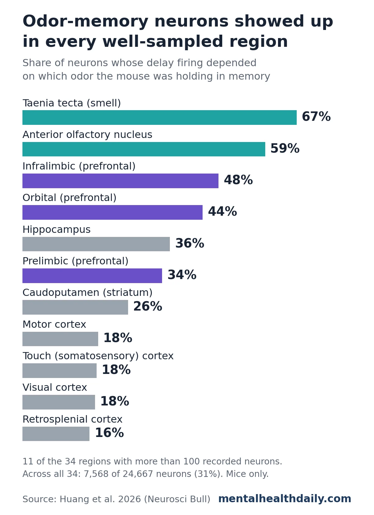
Remarkably, every single one of the 34 brain regions that were sufficiently sampled (i.e., had more than 100 recorded neurons) contained a significant proportion of these memory-content-selective cells. This discovery fundamentally challenges the long-held notion of working memory being primarily localized to a few specific "memory centers." While some regions exhibited a higher concentration of these neurons, the widespread distribution suggests a highly integrated and distributed neural network underpinning working memory.
As might be intuitively expected, regions heavily involved in smell processing and the prefrontal cortex — a region historically linked to executive functions and working memory — demonstrated the highest shares of memory-selective neurons. For instance, the taenia tecta, an olfactory-related area, showed a remarkable 67% of its neurons being memory-selective. Similarly, the anterior olfactory nucleus had 59%, the infralimbic cortex (part of the prefrontal cortex) 48%, and the orbital cortex 44%. The hippocampus, a structure critically involved in long-term memory formation, also showed a substantial 36% of its neurons being memory-selective, highlighting its potential role in short-term information retention as well.
Even areas traditionally associated with sensory processing (visual, somatosensory) and motor control, which are typically not considered primary sites for working memory storage, exhibited a discernible presence of these memory-selective neurons, albeit with smaller shares (e.g., motor cortex 18%, somatosensory cortex 18%, visual cortex 18%, and retrosplenial cortex 16%). This broad distribution suggests a more holistic brain engagement in maintaining information.
Beyond percentages, the sheer numbers of selective neurons in some regions were considerable. The orbital area alone contained 1,091 memory-selective neurons, the caudoputamen (a part of the striatum implicated in movement and reward learning) hosted 902, and the hippocampus contributed 670. These large absolute counts further underscore the significant involvement of these diverse brain regions.
A crucial aspect of the study was to confirm that the observed delay activity was indeed related to memory and not merely a prolonged sensory response to the odor. The researchers meticulously measured that the physical odor itself cleared from the environment to under 1% of its peak concentration within just one second of the odor valve closing. This rapid clearance definitively indicates that the sustained neural firing during the 3-6 second delay was not due to the smell physically lingering in the air but rather reflected an internal neural representation of the remembered odor. While some lingering sensory processing within neurons in smell-related areas could still contribute to delay firing, the dataset provides an excellent platform for future studies to disentangle pure sensory persistence from genuine memory maintenance. It’s also important to note that the reported percentages are descriptive counts from the dataset and do not represent formal statistical comparisons between regions, meaning the lowest shares are less definitive than the overall pattern of widespread distribution.
Reinforcing a Paradigm Shift in Neuroscience
The findings from this mouse dataset strongly align with a growing body of evidence that challenges the traditional modular view of brain function, particularly concerning complex cognitive processes like working memory. For decades, neuroscience often sought to pinpoint specific brain "centers" for particular functions. However, a 2017 review analyzing human and monkey studies on working memory already suggested a more distributed model, finding that activity carrying remembered information appeared not just in the prefrontal cortex, but also in sensory and parietal cortical areas.
This emerging understanding is further supported by other large-scale Neuropixels surveys in mice focusing on different behaviors. For example, studies on visual decision-making tasks and auditory processing have also demonstrated that neural signals relevant to these behaviors are widely distributed across various brain regions, rather than being concentrated in a single processing hub. However, a key distinction of the current dataset is its specific focus on a task involving a multi-second memory delay, thereby filling a critical gap in the existing literature by providing a brain-wide recording set explicitly designed to investigate the neural underpinnings of information maintenance over time. This continuous re-evaluation of how the brain processes information underscores a broader paradigm shift in neuroscience towards understanding brain function as an emergent property of dynamic, distributed networks rather than isolated modules.
The Transformative Power of Open Science: A Catalyst for Discovery
The release of this massive dataset in the Neurodata Without Borders (NWB) format is as significant as the scientific findings themselves. It embodies the principles of open science, promoting transparency, reproducibility, and collaborative discovery. By making this rich resource freely available, the researchers are inviting the global scientific community to build upon their work, validate their findings, and pose entirely new questions that might not have been conceived by the original team.
Indeed, the same recordings served as the foundation for a complementary 2025 study by the same research group, which has already yielded further insights. That study revealed that neurons within and across different brain regions coupled their spikes within milliseconds, and this precise temporal coupling was particularly concentrated among memory-selective neurons that preferred the same odor. Furthermore, these coupled spikes formed intricate, repeating activity patterns. These patterns were observed to replay not only during the task but also before and after trials, and even during the inter-trial breaks. Intriguingly, these patterns were organized in hierarchical layers, with larger, more complex patterns lasting longer and involving a greater number of neurons, suggesting a sophisticated organizational principle for memory representation.
With the data now public, other research groups are empowered to:
- Replicate and Validate: Independently verify the findings from the 2025 study and the current data paper, strengthening the robustness of the scientific conclusions.
- Investigate Memory Maintenance Mechanisms: Explore how information is precisely maintained across seconds within these distributed networks, potentially identifying novel neural codes or computational strategies.
- Characterize Cell-Type Specific Roles: Examine how different types of neurons (e.g., excitatory vs. inhibitory, specific genetic markers) contribute to working memory in various brain regions.
- Analyze Cross-Regional Communication: Understand how memory-related information is transmitted and integrated across different brain areas, revealing the dynamic interplay between distant circuits.
- Explore Brain States and Memory: Investigate how different brain states (e.g., attention, arousal, motivation) influence the encoding and retrieval of working memories.
- Develop Novel Computational Models: Use the rich empirical data to build and test more accurate computational models of working memory, bridging the gap between theory and observation.
This open data philosophy fosters a more inclusive and efficient scientific ecosystem, allowing researchers without access to specialized equipment or large animal facilities to contribute meaningfully to neuroscience.
Bridging the Gap: Implications for Mental Health and Neurological Disorders
The fundamental understanding of working memory derived from studies like this holds profound implications for mental health. As previously noted, working memory problems are a highly consistent and debilitating feature of schizophrenia. A comprehensive 2005 meta-analysis, for instance, revealed significant deficits across a wide array of working memory tasks in individuals with schizophrenia, irrespective of whether the information was verbal, visual, or spatial. These deficits severely impact daily functioning, affecting everything from medication adherence to social interactions and vocational success. Similar working memory impairments are observed in conditions like ADHD, where difficulty sustaining attention and manipulating information is central, and in neurodegenerative diseases such as Alzheimer’s, where working memory decline often precedes more widespread cognitive deterioration.
While basic mouse recordings cannot directly explain the complex human pathologies of these conditions, they provide an invaluable "detailed reference for how a healthy brain holds information over seconds." This foundational knowledge is crucial. Future studies can leverage this healthy baseline to:
- Model Disease States: Investigate working memory in mouse models of schizophrenia, ADHD, or Alzheimer’s disease to pinpoint specific neural circuit dysfunctions that lead to cognitive deficits.
- Identify Biomarkers: Discover unique neural signatures or patterns of activity that correlate with working memory impairments in disease models, potentially leading to diagnostic biomarkers.
- Develop Targeted Therapies: Test novel pharmacological or neuromodulatory interventions designed to restore or enhance working memory function, informed by a deep understanding of the healthy underlying circuitry.
- Compare Across Species: Use this detailed mouse data as a comparative framework for interpreting human brain imaging studies (e.g., fMRI, EEG), helping to translate findings between species and accelerate therapeutic development.
By elucidating the fundamental principles of how a healthy mammalian brain manages working memory, this dataset lays critical groundwork for understanding how these processes go awry in various neurological and psychiatric disorders, ultimately paving the way for more effective diagnostic tools and treatment strategies.
Limitations and Future Directions
Despite its immense scope and significance, it is crucial to acknowledge the inherent limitations of this dataset and the study design. Firstly, mice are not humans, and while their brains share many conserved features, there are significant anatomical and functional differences that necessitate caution when extrapolating findings directly to human cognition. The working memory task, while rigorously designed, is a simplified model of the complex, multifaceted working memory demands faced by humans in real-world scenarios. The head-fixed experimental setup, while necessary for stable neural recordings, also restricts the mouse’s natural behaviors and movements, which could potentially influence cognitive processes. Furthermore, the study focused on only two distinct odors, and future research will need to explore how the brain manages working memory for a broader range of sensory stimuli and increasing information load.
These limitations, however, are not drawbacks but rather define clear avenues for future research. The dataset itself serves as a springboard for further investigations, encouraging scientists to:
- Develop more complex working memory tasks for mice that incorporate higher cognitive demands or multimodal sensory information.
- Combine Neuropixels recordings with other techniques, such as optogenetics or chemogenetics, to causally manipulate specific neural circuits and assess their contribution to working memory.
- Conduct similar large-scale recordings in other model organisms or, where feasible, in humans, to draw more robust cross-species comparisons.
- Explore the long-term plasticity of these distributed memory networks and how they are modulated by learning and experience.
In conclusion, the release of this unprecedented dataset represents a pivotal moment in neuroscience. By demonstrating the brain-wide distribution of working memory-selective neurons in mice and providing an open-access window into this complex neural activity, Ermeng Huang and colleagues have not only advanced our fundamental understanding of a critical cognitive function but have also set a new standard for collaborative, data-driven discovery. This work underscores the power of cutting-edge technology combined with an open science ethos, promising to accelerate breakthroughs in our quest to understand the brain and address the challenges of mental health.

