

A recent study conducted by researchers at Mansoura University in Egypt has shed new light on the potential neuroprotective effects of losartan, a widely prescribed blood-pressure medication, in the context of toxin-induced brain injury. The investigation, which utilized a rat model with cerebellums damaged by carbon tetrachloride (CCl4), demonstrated that a four-week regimen of losartan significantly mitigated oxidative damage, inflammation, and tissue injury. Intriguingly, a nanoparticle formulation of losartan showed enhanced performance on several tissue and staining measures, though not consistently across all gene activity markers. These preliminary findings, published in a peer-reviewed journal, suggest a novel therapeutic avenue for addressing specific types of brain damage, particularly those involving the cerebellum.
Understanding the Vulnerable Cerebellum and the Threat of Toxin Exposure
The cerebellum, a fist-sized structure nestled at the back of the brain, plays a pivotal role far beyond its commonly recognized function in coordinating movement and maintaining balance. It is also intricately involved in cognitive processes, learning, and even emotional regulation. Its unique cellular architecture, particularly the presence of large and complex Purkinje cells, makes it exceptionally sensitive to various stressors, including toxins and the damaging effects of oxidative stress. Purkinje cells, the primary output neurons of the cerebellar cortex, are metabolically active and highly susceptible to neuronal degeneration, which can lead to severe motor deficits, ataxia, and cognitive impairment.
Chemical toxins, such as carbon tetrachloride (CCl4), represent a significant threat to brain health. Historically used as an industrial solvent, CCl4 is well-known for its potent hepatotoxic (liver-damaging) and nephrotoxic (kidney-damaging) properties. Its mechanism of toxicity involves its metabolic activation by cytochrome P450 enzymes, particularly in the liver, leading to the formation of highly reactive free radicals. These free radicals initiate a cascade of events, including lipid peroxidation, which damages cell membranes, disrupts cellular integrity, and triggers oxidative stress and inflammation. While CCl4’s primary targets are often the liver and kidneys, evidence suggests it can also induce oxidative stress and neurotoxicity in various brain regions, including the cerebellum, even after systemic exposure. For research purposes, CCl4 is a valuable tool for modeling acute oxidative injury and inflammation in experimental animals, providing a consistent and reproducible method to study neuroprotective strategies.
The Enigma of Necroptosis: A Programmed Form of Inflammatory Cell Death
Central to the study’s focus on cellular damage is the concept of necroptosis, a distinct form of programmed cell death that has garnered increasing attention in neuroscience. Unlike apoptosis, which is a "quiet" form of cell suicide designed to remove unwanted cells without triggering inflammation, necroptosis is inherently inflammatory. When cells undergo necroptosis, their membranes rupture, releasing intracellular contents into the extracellular space. These released components, known as damage-associated molecular patterns (DAMPs), alert the immune system and initiate a robust inflammatory response, often exacerbating tissue damage.
The molecular machinery of necroptosis is complex, involving a series of key proteins. Receptor-interacting protein kinase 1 (RIPK1) and RIPK3 are critical kinases that form a necrosome complex. Upon activation, RIPK3 phosphorylates mixed lineage kinase domain-like (MLKL), which then translocates to the cell membrane, oligomerizes, and forms pores, leading to cell lysis. Caspase-8, a protease typically associated with apoptosis, plays a crucial regulatory role by acting as a brake on the necroptotic pathway. When Caspase-8 is active, it cleaves RIPK1 and RIPK3, effectively steering cells away from necroptosis and towards apoptosis or survival. However, in conditions where Caspase-8 is inhibited or overwhelmed, the necroptotic pathway can be unleashed, contributing significantly to tissue injury, particularly in neurodegenerative diseases and ischemic injury. The rising interest in necroptosis stems from observations that its activation is implicated in postmortem brains of individuals with Alzheimer’s disease, correlating with disease stage and cognitive decline. Furthermore, blocking necroptosis has shown promise in reducing neuron loss in mouse models of Alzheimer’s, highlighting its potential as a therapeutic target for neuroprotection.
Losartan: A Repurposed Drug with Neuroprotective Potential
Losartan belongs to a class of medications known as angiotensin receptor blockers (ARBs). Its primary clinical indications are the treatment of hypertension (high blood pressure) and the protection of kidneys in patients with diabetes. Losartan exerts its effects by blocking the angiotensin II type 1 (AT1) receptor, thereby preventing angiotensin II, a potent vasoconstrictor and pro-inflammatory peptide, from binding and initiating its deleterious actions. Beyond its well-established cardiovascular and renal benefits, a growing body of research suggests that ARBs like losartan may possess pleiotropic effects, including anti-inflammatory, antioxidant, and neuroprotective properties that are independent of their blood pressure-lowering capabilities. This has made them attractive candidates for repurposing in neurological disorders where oxidative stress and inflammation are key pathological drivers. The current study represents the first investigation into losartan’s efficacy against direct chemical injury to the cerebellum, building upon existing evidence of its neuroprotective actions in other brain injury models.
The Experimental Design: A Detailed Look at the Rat Study
To investigate the neuroprotective potential of losartan, Nermeen Lashine et al. at Mansoura University meticulously designed an animal experiment. Twenty-four adult male Sprague-Dawley rats, a common strain in biomedical research known for its robust health and genetic uniformity, were randomly allocated into four distinct groups of six animals each:
- Control Group: These rats received no CCl4 and no treatment, serving as a baseline for normal cerebellar tissue.
- CCl4-Injured Group (Untreated): These rats received CCl4 to induce cerebellar damage but no subsequent losartan treatment, acting as the primary disease model comparator.
- Regular Losartan Group: These rats received CCl4 followed by treatment with standard losartan.
- Nanoparticle Losartan Group: These rats received CCl4 followed by treatment with the nanoparticle formulation of losartan.
The injury phase involved the administration of carbon tetrachloride (CCl4) to the rats in the three experimental groups. This was done to establish a clear model of cerebellar damage before any intervention. Critically, losartan treatment commenced only after the injury phase concluded, meaning the experiment was designed to assess whether losartan could repair or mitigate existing damage rather than prevent it. This distinction is crucial for understanding the therapeutic implications. The treatment period spanned four weeks, allowing sufficient time for the drug to exert its effects and for researchers to observe changes in tissue pathology.
A novel aspect of this study was the use of a nanoparticle version of losartan. These nanoparticles were prepared by grinding pure losartan in a ball mill, a mechanical process that reduces particle size to the nanoscale without the addition of carriers or coatings. The rationale behind using nanoparticles is rooted in pharmacokinetics: smaller particles possess a significantly larger surface area-to-volume ratio, which can dramatically alter a drug’s dissolution rate, absorption, and bioavailability. For brain-targeted therapies, nanoparticles can theoretically enhance drug penetration across the blood-brain barrier (BBB), although this was not explicitly tested here. The research team confirmed the spherical shape of their nanoparticles using electron microscopy. However, a notable limitation acknowledged by the authors was the absence of measurements for particle size distribution, drug levels in the blood or brain, or detailed pharmacokinetic studies to ascertain how these tiny crystals behaved within the body. These measurements are typically essential for fully characterizing nanoparticle drug delivery systems.
At the conclusion of the ten-week study period (six weeks for injury/recovery followed by four weeks of treatment), researchers harvested cerebellar tissue from all rats. A comprehensive battery of assessments was then performed to quantify the extent of damage and the efficacy of the treatments. These measures included:
- Oxidative Stress Markers: Levels of malondialdehyde (MDA), an indicator of lipid peroxidation and oxidative damage, were measured. The activity of key antioxidant enzymes such as superoxide dismutase (SOD), catalase (CAT), and glutathione (GSH) were also assessed to gauge the brain’s endogenous defense mechanisms.
- Inflammation Markers: The expression of tumor necrosis factor-alpha (TNF-alpha), a prominent pro-inflammatory cytokine, was quantified.
- Necroptosis Markers: Gene expression levels (messenger RNA) and protein staining for RIPK1, RIPK3, and MLKL were analyzed to monitor the activation status of the necroptosis pathway.
- Tissue Structure (Histopathology): Hematoxylin and Eosin (H&E) staining was used for general tissue morphology and identification of lesions. Nissl staining specifically visualizes neuronal cell bodies and can reveal neuronal degeneration or loss. Myelin staining assesses the integrity of myelin sheaths, crucial for nerve signal transmission. Glial fibrillary acidic protein (GFAP) staining, a marker for astrocytes, indicates astrogliosis, a common inflammatory response in brain injury.
- Blind Assessment: To ensure objectivity and minimize bias, investigators who scored the tissue slides were blinded to the group assignment of each sample.
The Devastating Impact of Carbon Tetrachloride on the Cerebellum
The results unequivocally demonstrated the severe neurotoxic effects of CCl4 on the rat cerebellum. Compared to the healthy control group, rats that received CCl4 but no subsequent treatment exhibited widespread and profound damage:
- Increased Oxidative Stress: Levels of malondialdehyde (MDA) were significantly elevated, indicating extensive lipid peroxidation. Concurrently, the activity of crucial antioxidant enzymes like superoxide dismutase (SOD), catalase (CAT), and glutathione (GSH) were markedly reduced, signaling a severe compromise of the cerebellum’s intrinsic defense against free radicals.
- Inflammation and Necroptosis Activation: There was a significant upregulation in the gene expression and protein levels of key necroptosis markers, including RIPK1, RIPK3, and MLKL. Additionally, levels of the pro-inflammatory cytokine TNF-alpha were elevated, confirming an active inflammatory process.
- Severe Tissue Injury: Histopathological examination revealed significant neuronal degeneration and loss, particularly affecting the sensitive Purkinje cells. Myelin staining showed extensive demyelination, indicative of damage to the white matter tracts essential for cerebellar function. GFAP staining revealed pronounced astrogliosis, a reactive proliferation of astrocytes, which is a hallmark of central nervous system injury and inflammation.
These findings are consistent with previous animal studies that have shown that even a single systemic dose of CCl4 can induce widespread lipid damage and deplete antioxidant enzymes across various brain regions, underscoring its potential as a neurotoxin.
Losartan’s Broad Protective Effects Against Cerebellar Damage
Against this backdrop of severe CCl4-induced injury, both the regular and nanoparticle formulations of losartan demonstrated significant neuroprotective effects. Compared to the untreated CCl4-injured rats, both treated groups showed substantial improvements across every category of measure: oxidative stress, gene activity, tissue structure, and protein staining.
Specifically, losartan treatment led to:
- Reduced Oxidative Stress: MDA levels were significantly lowered, and the activity of SOD, CAT, and GSH enzymes were partially restored, indicating a reduction in free radical damage and an enhancement of antioxidant defenses.
- Suppressed Inflammation: Levels of TNF-alpha were reduced, signifying a dampening of the inflammatory response.
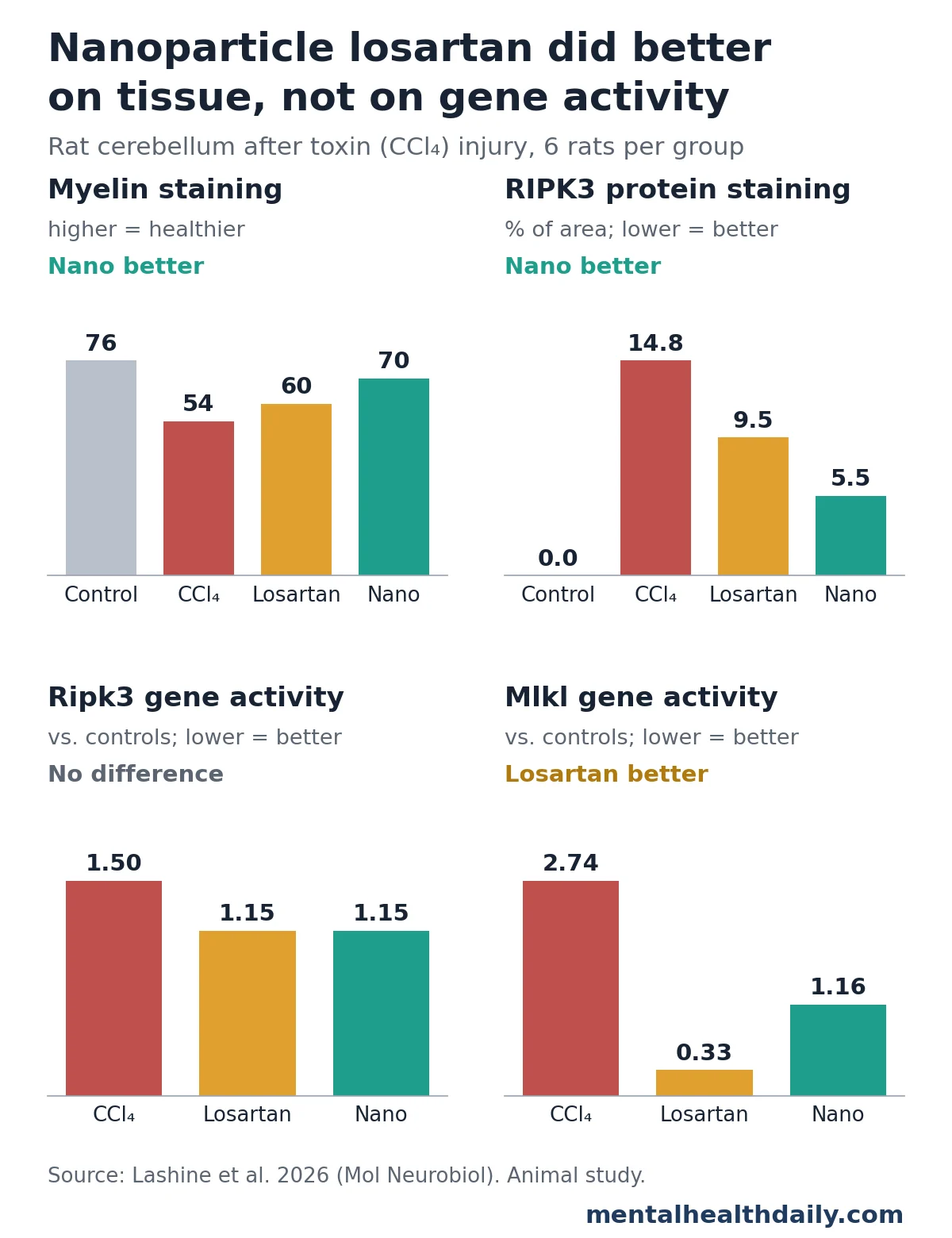
- Modulated Necroptosis Markers: Both gene expression and protein levels of RIPK1, RIPK3, and MLKL were significantly decreased. For instance, RIPK3 protein staining, which covered 14.8% of the tissue in the untreated CCl4 group, fell to 9.5% with regular losartan and 5.5% with nanoparticles, compared to 0% in controls. Similarly, Ripk3 gene activity, which was 1.50 after the toxin, dropped to 1.15 with both forms of losartan.
- Improved Tissue Integrity: Histopathological analysis revealed healthier neuronal morphology, increased Purkinje cell counts, reduced demyelination (e.g., myelin staining improved from 54.4% in CCl4 group to 60.5% with regular losartan and 69.5% with nanoparticle losartan, compared to 75.7% in controls), and decreased astrogliosis.
It is important to note that while improvements were significant, most measures did not return fully to the levels observed in the control group. For example, RIPK3 staining, though reduced, remained above the control baseline in both treated groups, suggesting a partial rather than complete reversal of the pathological processes.
Nanoparticle Losartan: A Mixed Bag of Advantages and Caveats
One of the study’s most intriguing aspects was the comparison between regular losartan and its nanoparticle counterpart. While the nanoparticle formulation did not consistently outperform the standard drug, it demonstrated a significant advantage in several key measures:
- Superior Tissue Staining and Cell Counts: Compared to regular losartan, the nanoparticle group exhibited significantly better results in myelin staining, indicating greater preservation of white matter integrity. They also showed healthier Nissl staining and higher Purkinje cell counts, suggesting better neuronal survival. Additionally, GFAP staining for astrogliosis was lower in the nanoparticle group, pointing to reduced inflammatory glial activation.
- Enhanced Necroptosis Protein Reduction: Nanoparticle losartan led to a more pronounced reduction in RIPK3 protein staining (5.5% vs. 9.5% for regular losartan), suggesting a greater impact on the necroptotic machinery at the protein level.
However, the picture was not uniformly superior for the nanoparticles, particularly concerning gene activity. Messenger RNA levels for genes like Tnf, Ripk1, and Ripk3 fell to similar levels with both forms of the drug. In a notable instance, Mlkl, the gene encoding the protein that directly ruptures the cell membrane during necroptosis, actually fell further with regular losartan (0.33) compared to the nanoparticle version (1.16). This discrepancy underscores the complexity of drug action at different biological levels and highlights the need for a more comprehensive understanding of the nanoparticles’ pharmacodynamics.
The researchers presented the observed nanoparticle advantage as an observation without drawing definitive conclusions about its superiority. This cautious interpretation is appropriate given the study’s limitations. Without data on how much of each drug formulation reached the brain, it is impossible to definitively determine whether the nanoparticle crystals were absorbed more effectively, exhibited different biological activity, or if the observed differences were merely due to chance variations within the small groups of six rats. Further studies with pharmacokinetic analyses are essential to elucidate the true benefits, if any, of the nanoparticle formulation.
The Necroptosis Pathway: Association, Not Definitive Activation
While the study meticulously measured levels of RIPK1, RIPK3, and MLKL, which are crucial components of the necroptosis pathway, the authors prudently clarified that these findings indicate an association with necroptosis-related changes rather than direct proof of pathway activation or blockade. Necroptosis is activated when RIPK3 and MLKL are phosphorylated – tagged with phosphate groups – which leads to their functional activation and subsequent cell membrane rupture. The research team did not measure these phosphorylated, activated forms of the proteins. Therefore, while the machinery for necroptosis was clearly more abundant and modulated by losartan, the study cannot definitively state that the pathway was "switched on" or "switched off" in a functional sense. The observed improvements could also stem from a broader reduction in oxidative stress and overall tissue injury, rather than a specific blockade of necroptosis. This distinction is vital for precisely understanding the mechanisms of neuroprotection.
Broader Context: Losartan and Brain Health
This study adds a crucial piece to the growing body of evidence supporting losartan’s neuroprotective potential. While this is reportedly the first test of losartan against chemical injury to the cerebellum, other animal studies have pointed in a similar direction:
- Ischemic Brain Injury: Losartan has been shown to reduce inflammation and oxidative stress in animal models of cerebral ischemia (stroke), suggesting its potential to mitigate damage caused by reduced blood flow to the brain.
- Traumatic Brain Injury: In models of traumatic brain injury, losartan has been observed to improve neurological outcomes and reduce neuronal death, possibly through its anti-inflammatory and anti-apoptotic effects.
- Alzheimer’s Disease Models: Some preclinical studies have indicated that losartan can reduce amyloid-beta plaque accumulation and improve cognitive function in mouse models of Alzheimer’s disease, linking it to broader neurodegenerative processes.
Beyond animal research, observational studies in humans have also hinted at a potential cognitive benefit. A large cohort study involving mostly male US veterans found that individuals taking angiotensin receptor blockers (ARBs), the class to which losartan belongs, developed dementia at lower rates compared to those on other heart medications (hazard ratio 0.76, indicating approximately a 24% lower risk) or even compared to those taking ACE inhibitors like lisinopril (0.81). While compelling, this type of observational study cannot establish a cause-and-effect relationship. It is impossible to fully separate the drugs’ effects from inherent differences in the health status or lifestyle of the individuals who were prescribed them. Furthermore, these findings pertain to long-term dementia risk in a general population, and have no direct bearing on acute toxin-induced brain injury, as modeled in the rat study. Nevertheless, such data provide a broader context for exploring the neuroprotective potential of ARBs.
Limitations and Future Directions
It is crucial to interpret the findings of this Losartan nanoparticle rat study within its limitations. As an early animal experiment, it can demonstrate that losartan altered tissue and marker measures after toxin injury in rats; it cannot, however, establish benefit for human patients or guarantee improvements in brain function. Key limitations include:
- Animal Model vs. Human Relevance: Findings in rats, while indicative, do not always translate directly to human physiology or disease. The CCl4 model, while useful for studying oxidative stress, does not represent a common human brain injury.
- Lack of Functional Outcomes: The study focused exclusively on biochemical and histological markers. It did not include behavioral tests (e.g., motor coordination, balance tests) that would provide direct evidence of improved brain function.
- Pharmacokinetic Data Gap: The absence of drug-level measurements in the blood and, critically, in the brain for both losartan formulations is a significant limitation, particularly for interpreting the nanoparticle advantage. Without knowing how much drug reached the target tissue, it’s difficult to conclude if nanoparticles enhanced delivery.
- Small Sample Size: With only six rats per group, the statistical power to detect subtle but real differences, especially between the two losartan formulations, may be limited.
- Short-Term Treatment: The four-week treatment period provides insights into acute recovery, but long-term effects and sustained neuroprotection remain unknown.
- Necroptosis Confirmation: The lack of measurement of phosphorylated RIPK3 and MLKL means the direct activation or inhibition of the necroptosis pathway was not conclusively proven.
For the science to advance, several next steps are warranted. Future research should include comprehensive pharmacokinetic studies to determine drug concentrations in the brain. Behavioral tests are essential to evaluate whether the observed biochemical and histological improvements translate into functional recovery. Longer-term studies are needed to assess sustained neuroprotection and potential side effects. Furthermore, investigating different dosages, exploring the specific cell types affected by losartan, and confirming the precise mechanisms of action, especially concerning necroptosis phosphorylation, would significantly strengthen the findings. Ultimately, if these preclinical results continue to be promising, eventual clinical trials in humans would be necessary, though likely for different types of neurological conditions than acute CCl4 exposure.
What This Means for Losartan and Brain Injury
From a scientific perspective, this study adds valuable evidence to the growing body of animal data suggesting that losartan can reduce oxidative stress and inflammation in brain tissue, now specifically in a cerebellar toxin model. It highlights the potential for repurposing existing drugs for novel therapeutic applications in neuroprotection. However, the question of whether grinding losartan into nanoparticles offers a true, reproducible advantage remains to be definitively answered, requiring more rigorous drug-level measurements and functional tests.
For the general public, it is crucial to understand that losartan is a prescription medication primarily indicated for managing high blood pressure and protecting kidney function. It is not currently approved or prescribed for brain injury or toxin exposure. Nanoparticle losartan, as explored in this study, is an experimental laboratory preparation and is not available for clinical use. Anyone who believes they have been exposed to solvents such as carbon tetrachloride, or who is experiencing balance, coordination problems, or other neurological symptoms, should seek immediate medical attention from a qualified healthcare professional. Self-treating with prescription medications based on preclinical animal research is ill-advised and potentially dangerous. This research offers a glimpse into future possibilities for neuroprotective therapies, but its translation to human clinical practice is a long and complex journey.

