

A comprehensive MRI study involving 337 young individuals aged 8 to 18 has unveiled distinct patterns of brain structural differences in autistic boys and girls when compared to their neurotypical peers of the same sex. The groundbreaking research, which leveraged advanced morphology-based connectivity analysis, indicates that these neurobiological distinctions may contribute to the diverse manifestations of autism spectrum disorder (ASD) across sexes. For autistic boys, the identified pattern of structural similarity centered on a critical face-processing region of the brain, exhibiting a direct correlation with social and communication traits. In contrast, autistic girls displayed a unique pattern localized around a key memory hub, a difference that notably did not correlate with their social and communication characteristics, suggesting varied underlying neural mechanisms.
This study, spearheaded by a team including Nooshin Safari and Javier Rasero at the University of Virginia, utilized standard structural MRI scans, which provide detailed anatomical snapshots of the brain rather than capturing its activity. The MRI data was sourced from the initial phase of the NIH Autism Centers of Excellence (ACE) Network, a crucial initiative recognized for its deliberate and balanced recruitment of equal numbers of autistic boys and girls. This balanced cohort is particularly significant given the historical male-centric bias prevalent in autism research. Traditionally, studies have featured a disproportionately higher number of male participants, largely due to autism being diagnosed roughly three to four times more frequently in males. However, meta-analyses, such as that conducted by Loomes et al., suggest that the true male-to-female ratio may be closer to 3:1, further indicating that girls who meet diagnostic criteria are often less likely to receive a formal diagnosis, a phenomenon sometimes attributed to "camouflaging" or differing presentation of traits.
Unpacking the Methodology: How Brain "Shape Networks" Were Constructed
The core of this research hinges on a sophisticated technique known as morphology-based connectivity. Unlike methods that track nerve fibers or brain activity, this approach assesses the structural likeness between different brain regions. The premise is that regions exhibiting greater physical similarity in their anatomical features are considered to have a stronger "connection" within this morphological network.
To establish these brain "shape networks," researchers meticulously analyzed 68 regions of the cortex, the brain’s intricately folded outer layer. For each of these regions, five distinct features were measured at thousands of points:
- Cortical thickness: The depth of the gray matter.
- Surface area: The extent of the cortical surface.
- Gray matter volume: The total volume of neuronal cell bodies.
- Mean curvature: A measure of the average bending of the cortical surface.
- Folding index: A quantification of how folded or convoluted a region is.
These detailed measurements formed a unique "structural fingerprint" for each cortical region. The study then employed a method called MIND (Morphometric Inverse Divergence) to compare the full spectrum of these measurements between every pair of regions. The more similar the structural fingerprints of two regions, the stronger their morphology-based connection was deemed to be. A crucial 2023 validation study, encompassing over 11,000 scans, underscored the reliability of MIND networks, demonstrating their superiority over older similarity methods and their better alignment with known nerve-fiber wiring, thereby bolstering the credibility of this analytical approach.
Following this intricate comparison, each participant was assigned a unique map detailing 2,278 region-to-region connections. Subsequently, a technique known as network-based statistics was utilized to identify clusters of interconnected regions that exhibited significant differences between the study groups, carefully adjusting for variables such as age, head size, and the specific MRI scan site to ensure robust results.
Distinct Patterns in Autistic Boys: A Focus on Face Processing
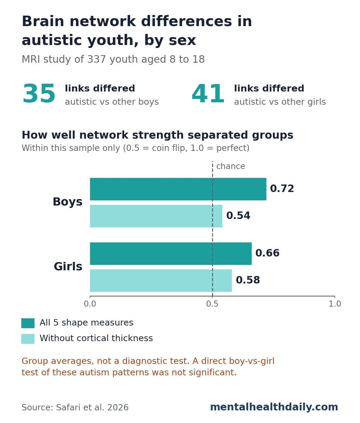
The study revealed a compelling pattern in autistic boys. When compared to a control group of 78 non-autistic boys, the 97 autistic boys in the sample displayed a distinct cluster of 35 connections that differed significantly. A striking observation was that nearly all these connections indicated regions being more structurally alike in the autistic group. Only one weak connection, situated between two temporal-lobe regions, showed the opposite trend.
Key regions implicated in the boys’ cluster included:
- Fusiform gyrus: Often referred to as the "fusiform face area," this region plays a crucial role in face perception, recognition, and social cognition. Differences in its activity and structure have long been associated with autism, particularly challenges in processing social cues from faces.
- Superior temporal sulcus (STS): Involved in processing biological motion, including eye gaze and mouth movements, which are vital for understanding social intentions and communication.
- Medial prefrontal cortex (mPFC): A central component of the brain’s "social brain network," involved in self-referential thought, theory of mind, and understanding the mental states of others.
Intriguingly, within the group of autistic boys, this specific network of structural connections demonstrated a significant correlation with social and communication scores derived from the ADI-R (Autism Diagnostic Interview-Revised), a standardized parent interview used to assess autism traits. The correlation was described as negative (r = 0.39), meaning that stronger morphology-based connectivity within this network was associated with less severe social-communication challenges. This finding suggests a potential compensatory mechanism or a spectrum of neurobiological organization related to the severity of these core autism traits in boys.
Autistic Girls: A Memory Hub and Uncharted Correlations
The findings for autistic girls presented a contrasting picture. Compared to 78 non-autistic girls, the 84 autistic girls in the study exhibited a cluster of 41 distinct connections. Similar to the boys’ group, most of these connections showed regions being more structurally alike in the autistic girls.
The primary region anchoring the girls’ unique pattern was the entorhinal cortex. This brain area is a critical component of the limbic system, acting as a major hub for memory formation and retrieval, particularly spatial navigation and episodic memory (memories of specific events). Its involvement in autism in girls opens new avenues for understanding how cognitive functions, beyond typical social-communication metrics, might be uniquely impacted or organized.
A key divergence from the findings in boys was the absence of a significant link between the girls’ distinct brain network and their autism trait scores. Furthermore, neither the boys’ nor the girls’ networks showed any significant correlation with scores related to anxiety, mood, attention, or behavior problems as reported on a parent checklist. This lack of correlation in girls, especially concerning their autism traits, underscores the potential for different pathways and manifestations of ASD across sexes, suggesting that the neurobiological underpinnings of social-communication challenges might be processed or compensated for differently in girls, or that other, as yet unidentified, behavioral or cognitive traits might correlate with these structural differences.
Are the Sex-Specific Patterns Statistically Different?
While the clusters of differing connections in autistic boys and girls appeared distinct, centering on different brain regions, the study’s formal statistical test for a sex-by-diagnosis interaction did not yield a significant result. This interaction test aims to determine whether the autism-related brain differences observed in boys were statistically distinct from those found in girls. Despite trying three different statistical cutoffs, no significant cluster emerged, leading the researchers to suggest that any true sex difference in these brain patterns might be too subtle to detect with the current sample size. This highlights a common challenge in neuroscience research: visual differences do not always translate to statistically significant distinctions, and larger, more diverse cohorts may be necessary to uncover such nuances.
It is important to note that these autism-related patterns were clearly distinguishable from ordinary sex differences observed in neurotypical individuals. The research team also mapped how non-autistic boys and girls differed from each other, identifying a 48-connection network. This "ordinary" sex difference network shared no connections with the boys’ autism cluster and only a single connection with the girls’ autism cluster, reinforcing that the identified patterns are specific to autism, not just general biological differences between sexes. Earlier preliminary work using a simpler, single-feature method on a younger cohort (2 to 4 years old) had reported sex-dependent network differences, but variations in age, methodology, and sample size preclude direct comparison between the studies.
Why These Brain Differences Are Not a Diagnostic Test
Despite the fascinating insights, it is crucial to emphasize that these observed brain differences are not, and should not be interpreted as, a diagnostic test for autism. The findings represent group-level averages, and there was substantial overlap between the autistic and non-autistic groups in terms of their brain network characteristics. Consequently, a single MRI scan cannot, based on this research, determine whether an individual child is autistic.
The researchers evaluated the ability of each network’s overall strength to differentiate between autistic and non-autistic youth within the study sample. The area under the curve (AUC), a measure where 0.5 indicates a random chance separation and 1.0 signifies perfect separation, was 0.72 for boys and 0.66 for girls. While these figures suggest a modest level of separation, it is vital to acknowledge that these AUC values were derived from the same individuals used to discover the networks, a practice that typically inflates the apparent accuracy. Crucially, these findings were not independently validated in a separate, external group, which is a standard requirement for assessing true diagnostic utility.
Furthermore, the study highlighted that cortical thickness was the primary driver of these differences. When cortical thickness was excluded from the analysis, the AUC values plummeted to 0.54 for boys and 0.58 for girls, barely exceeding chance. In contrast, removing surface area or volume had negligible impact. This underscores the specific role of cortical thickness in these morphological distinctions. The researchers themselves have explicitly stated that their approach does not measure diagnostic usefulness and advocate for future machine-learning studies that incorporate external testing to explore potential diagnostic applications. Ultimately, autism continues to be diagnosed through a comprehensive assessment of developmental history and behavioral observations, a process that remains unchanged by these neuroimaging findings.
Limitations and Strengths of the Study
As with any scientific endeavor, this MRI study comes with inherent limitations. The cross-sectional nature of the study means it captures brain structure at a single point in time, precluding conclusions about developmental trajectories or causal relationships. The specific age range (8 to 18) limits generalizability to younger children or adults with autism. While the study controlled for several confounds, other unmeasured variables could potentially influence brain structure. Additionally, the lack of correlation between the girls’ brain network and social-communication traits necessitates further investigation into what other behavioral or cognitive aspects might be linked to these structural differences.
However, the study also boasts significant strengths. A major advantage is the balanced sample, both by sex and diagnosis, a rarity in autism imaging research that has historically been skewed towards male participants. This careful balancing enhances the validity and generalizability of the sex-specific findings. The robustness of the identified clusters was further reinforced by their persistence even after rigorous adjustment for scan quality and the various scan sites involved in the data collection. Moreover, the finding concerning the fusiform gyrus in autistic boys aligns well with a long-standing body of research on face processing difficulties in autism, providing external validation for this particular aspect of the study.
Broader Impact and Future Directions
This study significantly enriches our understanding of the neurobiological heterogeneity within autism, particularly highlighting the importance of considering sex as a critical factor. By revealing distinct structural brain patterns in autistic boys and girls, the research moves beyond a monolithic view of ASD, paving the way for more nuanced and personalized approaches.
The implications for future research are profound. The findings underscore the urgent need for more balanced research cohorts in autism studies, emphasizing the necessity of including more female participants to fully capture the spectrum’s diversity. Longitudinal studies are crucial to track how these structural differences evolve over time and how they relate to developmental outcomes and the trajectory of autism traits. Integrating multimodal imaging techniques (e.g., combining structural MRI with functional MRI or diffusion tensor imaging) could provide a more holistic picture of brain organization and function. Furthermore, the findings lay groundwork for machine learning applications aimed at identifying neurobiological biomarkers that could, in the distant future, potentially assist in early identification or stratification of individuals for targeted interventions.
From a clinical perspective, while not a diagnostic tool, this research offers insights that could eventually inform the development of sex-specific therapeutic strategies. Understanding that the neurobiological underpinnings of autism may differ between boys and girls could lead to more tailored and effective interventions. For example, therapies targeting social-communication skills in boys might benefit from a deeper understanding of the fusiform gyrus network, while interventions for girls might need to consider the unique role of memory hubs like the entorhinal cortex. This nuanced understanding is vital for moving towards truly personalized medicine in autism care, acknowledging that "autism" is not a single entity but a spectrum of experiences shaped by diverse biological and environmental factors.

